Part 10: Arrhythmias
Fri Jul 31 2026
By B. Hassan
Normal cardiac function relies on the organized flow of electric impulses through the heart in a highly coordinated fashion. Abnormalities of the electric rhythm are known as arrhythmias (also termed dysrhythmias) and are among the most common clinical symptoms encountered. The presentation of arrhythmias range from common benign palpitations to severe symptoms of low cardiac output and death.
Abnormally slow heart rhythms are termed bradycardias (or bradyarrhythmias). Fast rhythms are known as tachycardias (or tachyarrhythmias). Tachycardias are further characterized as supraventricular when they involve the atrium or AV node and designated ventricular when they originate from the His–Purkinje system or ventricles.
Arrhythmias result from alterations of impulse formation, impulse propagation, or both. While studying this part, it is important to recall that cardiac tissue is composed of cells that are electrically coupled and operate as a syncytium. As myocytes depolarize and current result in individual action potentials, the electrical activity rapidly propagates from one cell to the next with minimal resistance, spreading through a large mass of tissue.
The study of arrhythmias is fun and easy, however, the mechanisms and distinctions can be nuanced. I recommend taking ample break time, especially when you are stuck and reread the article multiple times.
Normal impulse formation
As described in part 2, electric impulse formation in the heart arises from the intrinsic automaticity of specialized cardiac cells. Although atrial and ventricular myocytes do not exhibit automaticity under normal conditions, the cells of the specialized conducting system do possess natural automaticity and are therefore termed pacemaker cells. The specialized conducting system includes the SA node, the AV node, and the ventricular conducting system. The latter is composed of the bundle of His, the bundle branches, and the Purkinje fibers. In pathologic states, myocardial cells outside the conducting system may also acquire automaticity.
The distinct populations of automatic cells in the electro-conduction pathway have different intrinsic rates of firing. These rates are determined by three variables that influence how fast the membrane potential reaches the firing threshold: (1) the rate (i.e. slope) of phase 4 spontaneous depolarization, (2) the maximum negative diastolic potential, and (3) the firing threshold.
A more negative maximum diastolic potential, or a less negative firing threshold, slows the rate of impulse initiation because it takes longer to reach the threshold value. Conversely, the greater the current generated by the funny (pacemaker) sodium channels, the steeper the slope of phase 4 and the faster the cell depolarizes. The size of the pacemaker current depends on the number and opening kinetics of the individual funny (pacemaker) channels through which this current flows.
Since all healthy myocardial cells are electrically connected by gap junctions, an action potential generated in one part of the myocardium will ultimately spread to other regions. When an impulse arrives at a cell that is not yet close to threshold, current from the depolarized cell will bring the adjacent cell’s membrane potential to the firing threshold. Thus, the pacemaker cells with the fastest rate of depolarization set the heart rate.
In the normal heart, the dominant pacemaker is the SA node, which at rest initiates impulses at a rate of 60 to 100 bpm. Because the SA node rate is faster than that of other tissues that possess automaticity, its repeated discharges prevent spontaneous firing of other potential pacemaker sites.
The SA node is known as the native pacemaker. Other cells within the specialized conduction system harbor the potential to act as pacemakers if necessary and are therefore called latent pacemakers (or ectopic pacemakers). In contrast to the SA node, the AV node and the bundle of His have intrinsic ring rates of 50 to 60 bpm, and cells of the Purkinje system have rates of approximately 30 to 40 bpm. These latent sites may initiate impulses and take over the pacemaker function if the SA node slows or fails to fire or if conduction abnormalities block the normal wave of depolarization from reaching them.
Override suppression
Not only does the cell population with the fastest intrinsic rhythm preempt all other automatic cells from spontaneously firing, but it also directly suppresses their automaticity. This phenomenon is called overdrive suppression. Cells maintain their transmembrane ion distributions because of the continuous activity of the Na+/K+-ATPase. Because its net transport effect is one positive charge in the outward direction, Na+/K+-ATPase creates a hyperpolarizing current (i.e. it tends to make the inside of the cell more negative). As the cell potential becomes more negative, additional time is required for spontaneous phase 4 depolarization to reach the firing threshold, and therefore, the rate of spontaneous firing is decreased.
Although the hyperpolarizing current moves the membrane voltage away from threshold, pacemaker cells have a pacemaker current sufficiently large to overcome this hyperpolarizing influence.
The more often the cell is depolarized, the greater the quantity of Na+ ions that enter the cell per unit time. As a result, Na+/K+-ATPase becomes more active, tending to restore the normal transmembrane Na+ gradient. This increased pump activity provides a larger hyperpolarizing current, opposing the depolarizing pacemaker current, and further decreases the rate of spontaneous depolarization. Thus, overdrive suppression decreases a cell’s automaticity when that cell is driven to depolarize faster than its intrinsic discharge rate.
Electrotonic Interactions
In addition to overdrive suppression, anatomic connections between pacemaker and non-pacemaker cells are important in determining how adjacent cells suppress latent pacemaker impulses.
Myocardial cells in the ventricle and Purkinje system repolarize to a resting potential of approximately −90 mV, whereas pacemaker cells in the SA and AV nodes repolarize to a maximum diastolic potential of about −60 mV. When these two cell types are adjacent to one another, they are electrically coupled through low-resistance gap junctions concentrated in their intercalated discs. This coupling results in a compromise of electric potentials owing to electrotonic current flow between the cells, causing relative hyperpolarization of the pacemaker cell and relative depolarization of the non-pacemaker cell. Hyperpolarization moves the diastolic potential further from threshold and thus slows the heart rate.
Electrotonic effects are particularly important in suppressing automaticity in the AV node (via connections between atrial myocytes and AV nodal cells) and in the distal Purkinje fibers (which are coupled to ventricular myocardial cells). In contrast, cells in the SA node are less tightly coupled to atrial myocytes; thus, their automaticity is less subject to electrotonic interactions.
Decoupling of normally suppressed cells, such as those in the AV node (e.g. by ischemic damage), may reduce the inhibitory electrotonic influence and enhance automaticity, producing ectopic rhythms by the latent pacemaker tissue.
Altered impulse formation
The main abnormalities of impulse initiation that lead to arrhythmias are altered automaticity, abnormal automaticity, and triggered activity.
Alterations in SA Node Automaticity
The rate of impulse initiation by the SA node, as well as by latent pacemakers is regulated primarily by neurohumoral factors. The most important modulator of normal SA node automaticity is the autonomic nervous system. Sympathetic stimulation through β1-adrenergic receptors increases the opening probability of the pacemaker channels, increasing the slope of phase 4 depolarization, causing the SA node to reach threshold and fire earlier than normal and the heart rate to increase.
In addition, sympathetic stimulation shifts the firing threshold to more negative voltages by increasing the probability that voltage-sensitive channels will opening (recall that calcium carries the current of phase 0 depolarization in pacemaker cells). Thus, phase 4 depolarization reaches the threshold potential earlier.
Normal decreases in SA node automaticity are mediated by reduced sympathetic stimulation and by increased activity of the parasympathetic nervous system. Whereas activation of the sympathetic nervous system has a major role in increasing the heart rate during times of stress, the parasympathetic nervous system is the major controller of the heart rate at rest.
Cholinergic (i.e. parasympathetic) stimulation via the vagus nerve acts at the SA node to reduce the probability of pacemaker channels being open, reducing the slope of phase 4 depolarization. In addition, the probability of the channels being open is decreased, such that the firing threshold increases to a less negative potential. Furthermore, cholinergic stimulation increases the probability of acetylcholine-sensitive K+ channels (different from the K+ channels active in phase 3 repolarization) being open at rest, causing a positive K+ outflow and cell hyperpolarization, slowing the intrinsic firing rate and reducing heart rate.
Escape rhythms escape
If the SA node becomes suppressed and fires much less frequently than normal, the site of impulse formation may shift to a latent pacemaker. An impulse initiated by a latent pacemaker because the SA node rate has slowed is called an escape beat.
Persistent impairment of the SA node allows a continued series of escape beats, termed an escape rhythm. Escape rhythms are protective in that they prevent the heart rate from becoming pathologically slow when SA node firing is impaired.
Different regions of the heart have varied sensitivities to parasympathetic (vagal) stimulation. The SA node and the AV node are most sensitive, while the ventricular conducting system is the least sensitive (vagal escape phenomenon). Therefore, moderate parasympathetic stimulation slows the sinus rate and allows the pacemaker to shift to the AV node. However, very strong parasympathetic stimulation suppresses excitability at both the SA node and AV node and may therefore result in the emergence of a ventricular escape pacemaker.
Ectopic firing foci
Another means by which a latent pacemaker can assume control of impulse formation is if it develops an intrinsic rate of depolarization faster than that of the SA node. Termed an ectopic beat, such impulse is premature relative to the normal rhythm, whereas an escape beat is late and terminates a pause caused by a slowed sinus rhythm. A sequence of ectopic beats is called an ectopic rhythm.
Ectopic beats may arise in several circumstances. For example, high catecholamine concentrations can enhance the automaticity of latent pacemakers, and if the resulting rate of depolarization exceeds that of the SA node, then an ectopic rhythm will develop. Ectopic beats are also commonly induced by hypoxemia, ischemia, electrolyte disturbances, and certain drug toxicities (e.g. digitalis).
Cardiac tissue injury may lead to pathologic changes in impulse formation whereby myocardial cells outside the electro-conduction system acquire automaticity. Although such activity may appear similar to impulses originating from latent pacemakers in the specialized conduction pathways, these ectopic beats arise from cells that do not usually possess automaticity. If the rate of depolarization of such cells exceeds that of the SA node, they transiently take over the pacemaker function and become the source of an abnormal ectopic rhythm.
Because these myocardial cells have few or no activated pacemaker channels, they do not normally exhibit pacemaker potential. How injury allows such cells to spontaneously depolarize is not fully understood. However, when cardiac tissue gets injured, its cell membranes become “leaky.” As such, they are unable to maintain the concentration gradients of ions, and the resting potential becomes less negative. When a cell’s resting membrane potential is reduced to a value less negative than −60 mV, gradual phase 4 depolarization can be demonstrated even among non-pacemaker cells.
Afterdepolarizations
Under certain conditions, an action potential can “trigger” abnormal depolarizations that result in extra heart beats or tachyarrhythmias. This process may occur when the first action potential leads to oscillations of the membrane voltage known as afterdepolarizations. Unlike the spontaneous activity seen when enhanced automaticity occurs, this type of automaticity is stimulated by a preceding action potential.
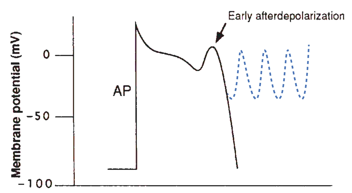
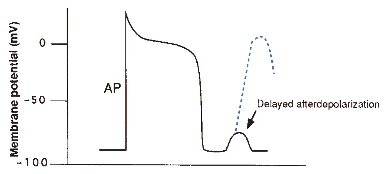
There are two types of afterdepolarizations depending on their timing after the inciting action potential: early afterdepolarizations occur during the repolarization phase of the inciting beat, whereas delayed afterdepolarizations occur shortly after repolarization has been completed.
Early afterdepolarizations are changes of the membrane potential that interrupt normal repolarization. They can occur either during the plateau of the action potential (phase 2) or during rapid repolarization (phase 3). Early afterdepolarizations are more likely to develop in conditions that prolong the action potential duration (i.e. the QT interval of the ECG), as may occur during therapy with certain drugs and in the inherited long QT syndromes.
The ionic current responsible for an early afterdepolarization depends on the membrane voltage at which the triggered event occurs. If the early afterdepolarization occurs during phase 2 of the action potential when most of the Na+ channels are still in an inactivated state, the upstroke of the triggered beat relies mostly on an inward current. If, however, the afterdepolarization occurs during phase 3, there is partial recovery of the inactivated Na+ channels, which then contribute more to the current underlying the triggered beat. An early afterdepolarization-triggered action potential can be self-perpetuating and lead to a series of depolarizations causing tachyarrhythmia. Early afterdepolarizations appear to be the initiating mechanism of the polymorphic ventricular tachycardia known as torsades de pointes.
Delayed afterdepolarizations may appear shortly after repolarization. They most commonly develop in states of high intracellular calcium, as in digitalis intoxication, or during marked catecholamine stimulation. It is thought that intracellular accumulation causes the activation of chloride currents, or of the Na+/Ca++ exchanger, resulting in brief inward currents that generate the delayed afterdepolarization. As with early afterdepolarizations, if the amplitude of the delayed afterdepolarization reaches a threshold voltage, an action potential is generated. Such action potential can be self-perpetuating, leading to tachyarrhythmias.
Altered impulse conduction
Alterations in impulse conduction also lead to arrhythmias. Conduction blocks generally slow the heart rate (bradyarrhythmias); however, under certain circumstances, the process of reentry can produce tachyarrhythmias.
Conduction block
A propagating impulse is blocked when it encounters a region of the heart that is electrically unexcitable. Conduction block can be either transient or permanent and may be unidirectional or bidirectional.
Various conditions may cause conduction block, including ischemia, fibrosis, inflammation, and certain drugs. When conduction block occurs because a propagating impulse encounters cardiac cells that are still refractory from a previous depolarization, the block is said to be functional. For example, antiarrhythmic drugs that prolong the action potential duration tend to produce functional conduction blocks. Conversely, when conduction block is caused by a barrier imposed by fibrosis, the block is said to be fixed.
Conduction block within the AV node or the His–Purkinje system prevents normal propagation of the cardiac impulse from the SA node to more distal sites. This AV block removes the normal overdrive suppression that keeps latent pacemakers in the His–Purkinje system in check. Thus, conduction block usually results in emergence of escape beats or escape rhythms, as the more distal sites resume their automaticity.
Reentry
A common mechanism by which altered impulse conduction leads to tachyarrhythmias is reentry. During such rhythm, an electric impulse circulates repeatedly around an abnormal path, recurrently depolarizing a region of cardiac tissue.
During normal cardiac conduction, each electric impulse that originates in the SA node travels in an orderly, sequential fashion, ultimately depolarizing all myocardial fibers. The refractory period of each cell prevents immediate re-excitation from adjacent depolarized cells, so that the impulse stops when all of the heart muscle has been excited. However, conduction blocks that prevent rapid depolarization of parts of the myocardium can create an environment conducive to continued impulse propagation and reentry.
The following figure shows electric activity as it flows through a branch point anywhere within the conduction pathways. Panel A shows propagation of a normal action potential. At point x, the impulse branches into two pathways (α and β). In the normal heart, the α and β pathways have similar conduction velocities and refractory periods such that impulses that pass through them may collide in the distal conduction tissue and cancel each other, as shown by the red line.
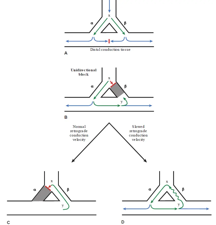
Panel B shows what happens if conduction is blocked in one of pathways. In this example, the action potential is obstructed when it encounters the β pathway and therefore propagates only down the α tract. As the impulse continues to spread, it encounters the distal end of the β pathway (point y). If the tissue in the distal β tract is also unable to conduct, the impulse simply continues to propagate into the deeper tissues and reentry does not occur. However, if the impulse at y is able to propagate retrogradely into pathway β, one of the necessary conditions for reentry is met.
When an action potential can conduct in a retrograde direction, whereas it had been prevented from doing so in the forward direction, unidirectional block is said to be present. Unidirectional block tends to occur in regions where the refractory periods of adjacent cells are heterogeneous, such that some cells recover before others. In addition, unidirectional block may occur in states of cellular dysfunction and in regions of fibrosis.
As shown in panel C, if the impulse is able to propagate retrogradely up the β pathway, it will again arrive at point x. At that time, if the α pathway has not yet repolarized from the previous action potential that had occurred moments earlier, that pathway is refractory to repeat stimulation and the returning impulse simply stops there. However, panel D illustrates what happens if the velocity of retrograde conduction in the diseased β path is slower than normal. In that case, sufficient time may elapse for the α pathway to repolarize before the returning impulse reaches point x from the β limb. Then, the invading impulse is able to stimulate the α pathway once again, and the cycle repeats itself. This circular stimulation can continue indefinitely resulting in a tachyarrhythmia.
For reentry to occur, the propagating impulse must continuously encounter excitable tissue. Thus, the time it takes for the impulse to travel around the reentrant loop must be greater than the time required for recovery (i.e. the refractory period) of the tissue, and this must be true for each point in the circuit. If the conduction time is shorter than the recovery time, the impulse will encounter refractory tissue and the loop stops.
Because normal conduction velocity in ventricular muscle is approximately 50 cm/s and the average effective refractory period is about 0.2 seconds, a reentry circuit would need to be at least 10 cm long for reentry to occur in a normal ventricle. However, with slower conduction velocities, a shorter reentry circuit is possible. Most clinical cases of reentry occur within small regions because the conduction velocity within the reentrant loop is abnormally slow.
In summary, the two critical conditions or reentry are unidirectional block and (2) slowed conduction through the reentry path. These conditions commonly occur in regions of fibrosis such as infarction scars. In some cases, reentry occurs over an anatomically fixed circuit using an accessory pathway.
Note
Reentry around distinct anatomic pathways usually appears as a monomorphic tachycardia on ECG; that is, in case of ventricular tachycardia, each QRS has the same appearance as the preceding and subsequent QRS complexes. This is because the reentry path is the same from beat to beat, producing a stable, regular tachycardia.
Other types of reentry do not require a stable, fixed path. For example, one form can occur in electrically heterogeneous myocardium, in which waves of reentrant excitation spiral through the tissue, continually changing direction. These “spiral waves” can be initiated when a wave of depolarization encounters a broad region of functional block, and forward wave propagation is asymmetrically blocked by this region, as the remainder of the wave proceeds around the block. As the region repolarizes and becomes excitable again, parts of the wave then spread retrogradely through it and continue in a spiral path. Unlike an anatomically fixed reentrant tract, the center of the spiral wave can move through the myocardium and even split into two or more reentry waves. In the ventricles, the resulting tachycardia has a continually changing QRS appearance, producing polymorphic ventricular tachycardia.
Wolff–Parkinson–White Syndrome
One of the most notable examples of reentry is the Wolff–Parkinson–White (WPW) syndrome. In the normal heart, an impulse generated by the SA node propagates through atrial tissue to the AV node, where physiological slower conduction causes a short delay before continuing to the ventricles. However, approximately 1 in 1,500 people has the WPW syndrome and are born with an additional conductive pathway between the atrium and ventricle. The most common type of accessory pathway consists of microscopic fibers (known as a bundle of Kent) that span the AV groove somewhere along the mitral or tricuspid annuli, and it allows conduction between the atria and ventricles to bypass the AV node.
Because the accessory pathway conducts impulses faster than the AV node, stimulation of the ventricles during sinus rhythm begins earlier than normal and the PR interval of the ECG is shortened (usually seconds, or small boxes). In this situation, the ventricles are said to be “preexcited.” However, the accessory pathway connects to ventricular myocardium rather than to the Purkinje system, such that the spread of the impulse through the ventricles from that site is slower than usual.
In addition, because normal conduction over the AV node proceeds concurrently, ventricular depolarization represents a combination of the electric impulse traveling via the accessory tract and that conducted through the normal Purkinje system. As a result, the QRS complex in these patients is wider than normal and demonstrates an abnormally slurred initial upstroke, known as a delta wave.
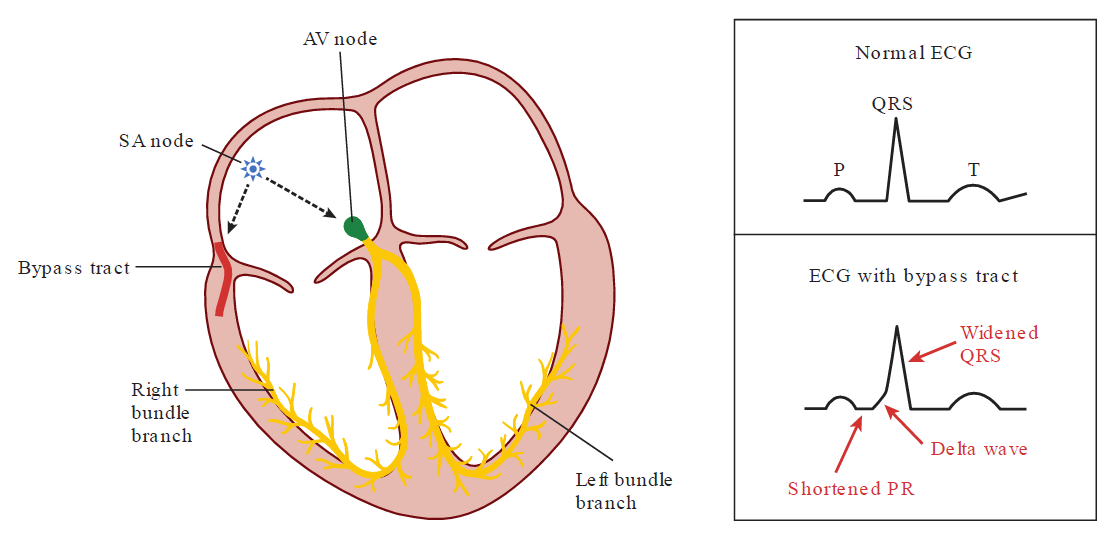
During sinus rhythm, simultaneous conduction through the accessory pathway and AV node results in this interesting ECG appearance but causes no symptoms. The presence of the abnormal pathway, however, creates an ideal condition for reentry because the refractory period of the pathway is usually different from that of the AV node. An appropriately timed abnormal impulse may encounter block in the accessory pathway but conduct through the AV node or vice versa. If the propagating impulse then finds that the initially blocked pathway has recovered (unidirectional block), it can conduct in a retrograde direction up to the atrium and then down the other pathway back to the ventricles. Thus, a large anatomic loop is established, with the accessory pathway serving as one limb and the normal conduction pathway through the AV node as the other.
Physiological basis of antiarrhythmic therapy
Appropriate treatment of arrhythmia depends on its severity and its likely mechanism. When an arrhythmia produces severe hypotension or cardiac arrest, it must be immediately terminated to restore effective cardiac function. Therapy for termination may include electrical cardioversion for tachycardias, cardiac pacing for bradycardias, or administration of medications.
Additional therapy to prevent recurrences is guided by the etiology of the rhythm disturbance. Correctable factors that contribute to abnormal impulse formation and conduction (such as ischemia or electrolyte abnormalities) should be corrected. If there is a risk of recurrent arrhythmia, medications that alter automaticity, conduction, and/or refractoriness may be administered, or catheter or surgical ablation of conduction pathways is undertaken to physically disrupt the region responsible for the arrhythmia.
Other advanced options include implantation of a permanent pacemaker for serious bradyarrhythmias or an internal cardioverter–defibrillator (ICD) to automatically terminate malignant tachyarrhythmias should they recur.
Bradyarrhythmias
Not all slow heart rhythms require specific treatment. For those that do, pharmacologic therapy can increase the heart rate acutely, but the effect is transient. Electronic pacemakers are used when sustained therapy is needed.
Pharmacologic therapy modifies the autonomic input to the heart in one of two ways:
-
Anticholinergic drugs (i.e. antimuscarinic agents such as atropine) as we talked about earlier
-
β1-Receptor agonists (e.g. isoproterenol). Mimicking the effect of endogenous catecholamines, these drugs increase heart rate and speed AV nodal conduction.
Atropine and isoproterenol are administered intravenously. Although these are useful in managing certain bradyarrhythmias emergently, it is not practical to continue long-term to treat persistent bradyarrhythmias.
Electronic pacemakers apply repeated electric stimulation to the heart to initiate depolarizations at a desired rate, thereby assuming control of the rhythm. Pacemakers may be installed on a temporary or a permanent basis. Temporary units are used to stabilize patients who are awaiting implantation of a permanent pacemaker or to treat transient bradyarrhythmias, such as those caused by reversible drug toxicities.
There are two types of temporary pacemakers. External transthoracic pacemakers deliver electric pulses to the patient’s chest through large adhesive electrodes placed on the skin. Unfortunately, because the current used must be sufficient to initiate a cardiac depolarization, it stimulates thoracic nerves and skeletal muscle, which can be uncomfortable. Therefore, this form of pacing is usually used only on an emergency basis until other means of treating the arrhythmia can be implemented.
The other option for temporary pacing is a transvenous unit. In this case, an electrode-tipped catheter is inserted percutaneously into the venous system, passed into the heart, and connected to an external power source (termed a pulse generator). Electric pulses are applied directly to the heart through the electrode catheter, which is typically placed in the right ventricle or right atrium. This type of pacing is not painful and can be effective for days. There is, however, a risk of catheter-associated infection and/or thrombosis.
Permanent pacemakers are more sophisticated. One or more wires (known as leads) with pacing electrodes are passed through an axillary or subclavian vein into the right ventricle or right atrium, or through the coronary sinus into a cardiac vein (to stimulate the left ventricle). The pulse generator, similar in size to two silver dollars stacked on top of one another, is connected to the leads and then implanted under the skin, typically in the infraclavicular region. The pacemaker battery typically lasts about 10 years. Modern permanent pacemakers sense cardiac activity and pace only when needed. They incorporate complex functions to track the patient’s normal heart rate and can stimulate beats automatically in response to activity. They can also record useful data, such as whether fast rates have been sensed (might indicate a tachyarrhythmia), the amount of pacing that has been required, and other parameters of pacemaker function. An external radio frequency programming device is used to “interrogate” the pacemaker to obtain the recorded information and to adjust the pacing functions.
Tachyarrhythmias
The treatment of tachyarrhythmias is directed at protection of the patient from the consequences of the arrhythmia and the treatment of the underlying cause responsible for the abnormal rhythm.
Pharmacologic management of tachyarrhythmias is directed against the underlying mechanism (abnormal automaticity, reentrant circuits, or triggered activity). From consideration of the arrhythmia mechanisms presented in this chapter, the following strategies emerge:
Desired drug effects to eliminate rhythms caused by increased automaticity:
-
Reduce the slope of phase 4 spontaneous depolarization of automatic cells
-
Make the diastolic potential more negative (hyperpolarize)
-
Make the threshold potential less negative
Desired Antiarrhythmic Effects to Interrupt Reentrant Circuits:
-
Inhibit conduction in the reentry circuit to the point that conduction fails, stopping the reentry impulse
-
Increase the refractory period within the reentrant circuit so that a propagating impulse finds tissue within the loop unexcitable and the impulse stops
-
Suppress premature beats that can initiate reentry
Desired drug effects to eliminate triggered activity:
-
Shorten the action potential duration (to prevent early afterdepolarizations)
-
Correct conditions of calcium overload (to prevent delayed afterdepolarizations)
Many drugs have multiple effects and may attack arrhythmias through more than one mechanism. It is important to recognize that although these drugs suppress arrhythmias, they also have the potential to provoke other rhythm disturbances. This undesired consequence is referred to as proarrhythmia and is a major limitation of contemporary antiarrhythmic drug therapy. For example, antiarrhythmic agents that prolong the action potential duration can cause early afterdepolarizations, the mechanism underlying the polymorphic ventricular tachycardia torsade de pointes. In addition, most agents used to treat tachyarrhythmias have the potential to aggravate bradyarrhythmias, and all antiarrhythmics have potentially toxic non-cardiac side effects. These shortcomings have led to an increased reliance on non-pharmacological treatment options.
Many tachycardias involve transmission of impulses through the AV node, a structure sensitive to vagal stimulation. Vagal tone can be transiently increased by a number of bedside maneuvers, which may slow conduction and terminate some reentrant tachyarrhythmias. For example, carotid sinus massage is performed by rubbing firmly for a few seconds over the carotid sinus, located at the bifurcation of the internal and external carotid arteries on either side of the neck. This maneuver stimulates the baroreceptor reflex, increasing vagal tone and decreasing sympathetic tone. This maneuver should be performed on only one carotid sinus at a time (to prevent interference with brain perfusion) and is best avoided in patients with known advanced atherosclerosis involving the carotid arteries.
Cardioversion and defibrillation involve the application of an electric shock to terminate a tachycardia. A shock with sufficient energy depolarizes the bulk of the myocardial tissue, interrupts reentrant circuits, establishes electric homogeneity, and allows the SA node to regain pacemaker control. Tachyarrhythmias that are caused by reentry can usually be terminated by this procedure, whereas arrhythmias due to abnormal automaticity simply persist.
External cardioversion is used to terminate supraventricular tachycardias or organized ventricular tachycardias. It is performed by briefly sedating the patient and then placing two large electrode paddles on either side of the heart. The electric discharge is synchronized to occur at the time of a QRS complex, which prevents the possibility of discharge during the T wave, when a shock could induce reentry (leading to ventricular fibrillation) because regions of myocardium are in different phases of depolarization and recovery.
External defibrillation is performed to terminate ventricular fibrillation, employing the same equipment used for cardioversion. However, because there are no organized QRS complexes during fibrillation, the device is used in the “asynchronous” mode.
ICDs automatically terminate dangerous ventricular arrhythmias using internal cardioversion/defibrillation or by way of a special type of artificial pacing. These devices are implanted in a manner similar to permanent pacemakers, in patients at high risk of sudden cardiac death from ventricular arrhythmias. The device continuously monitors cardiac activity, and if the heart rate exceeds a certain programmable threshold, the ICD delivers an appropriate intervention, such as an electric shock. Internal cardioversion or defibrillation requires substantially less energy than does external defibrillation but is still painful if the patient is conscious.
The majority of monomorphic ventricular tachycardias can be terminated by an ICD with a rapid burst of electric impulses, termed anti-tachycardia pacing or burst pacing, rather than a shock. The goal is to artificially pace the heart at a rate faster than the tachycardia to prematurely depolarize a portion of a reentrant circuit, thereby rendering it refractory to further immediate stimulation. An advantage of burst pacing is that, unlike cardioversion, it is painless. However, it is not effective for terminating ventricular fibrillation, a situation in which the device is programmed to deliver an electric shock instead.
If an arrhythmia originates from a distinct anatomical reentry circuit or an automatic focus, mapping techniques can be used to localize the region of responsible for the disturbance. It is then often possible to ablate that site via a catheter that applies radiofrequency current to heat and destroy the tissue. Such procedures have revolutionized the management of patients with many types of tachycardias because they often offer a permanent solution that spares patients from prolonged antiarrhythmic therapy.
Bradyarrhythmias
The normal resting heart rate, resulting from repetitive depolarization of the SA node, ranges from 60 to 100 bpm. Bradyarrhythmias are rhythms in which the heart rate is less than 60 bpm.
Sinus bradycardia
Sinus bradycardia is a slowing of the normal heart rhythm, as a result of decreased automaticity of the SA node, to a rate less than 60 bpm. Sinus bradycardia at rest or during sleep is a normal finding in many people. Conversely, pathologic sinus bradycardia can result from either intrinsic SA node disease or extrinsic factors that affect the node. Depressed intrinsic automaticity can be caused by aging or any disease that affects the atrium. Extrinsic factors that suppress SA nodal activity include medications (e.g. β-blockers and certain calcium channel blockers) and metabolic causes (e.g. hypothyroidism).
Trained athletes often have elevated vagal tone, resulting in physiologic asymptomatic resting sinus bradycardia. Transient periods of high vagal tone can also occur in individuals as a reflex to pain or fear.
Mild sinus bradycardia is usually asymptomatic and does not require treatment. However, a pronounced reduction of the heart rate can produce a fall in cardiac output with fatigue, light-headedness, confusion, or syncope. In such cases, any extrinsic provocative factors should be corrected, and specific therapy may be needed.
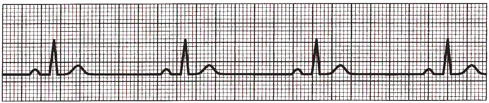
The following ECG tracing shows sinus bradycardia, with normal P waves and QRS complexes, but the rate is less than 60 bpm:
Sick Sinus Syndrome
Sick sinus syndrome (SSS) is intrinsic SA nodal dysfunction that causes periods of inappropriate bradycardia. It is often accompanied by symptoms of dizziness, confusion, or syncope, and mainly occurs in elder patients
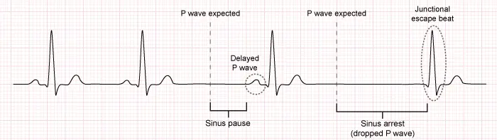
The following ECG trace shows SSS, with a sinus pause (delayed P wave) and a sinus arrest (dropped P wave) followed by a junctional escape beat.
Patients with this syndrome (or any symptomatic sinus bradycardia in general) can be treated acutely with IV anticholinergic drugs (e.g. atropine) or β-adrenergic agonists (e.g. isoproterenol), which transiently accelerate the heart rate. If the problem is chronic and not corrected by removal of aggravating factors, placement of a permanent pacemaker is required.
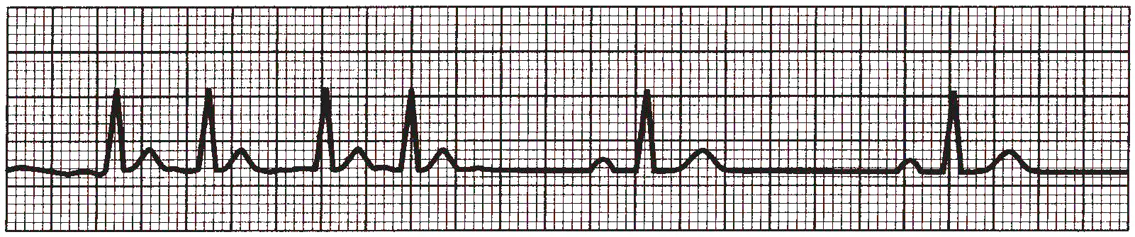
SSS is common in the elderly, who are also susceptible to supraventricular tachycardias (SVTs), most commonly atrial fibrillation (AF). This combination of slow and fast dysrhythmia is known as the bradycardia–tachycardia syndrome and is thought to result from atrial fibrosis that impairs function of the SA node and predisposes to AF and atrial flutter. During the tachyarrhythmia, overdrive suppression of the SA node occurs, and when the tachycardia terminates, a period of profound sinus bradycardia may ensue. Treatment generally requires the combination of antiarrhythmic drugs to suppress the tachyarrhythmias + a permanent pacemaker to prevent bradycardia.
The following ECG trace shows bradycardia tachycardia syndrome, with a period of tachycardia followed by a period of bradycardia.
Escape rhythm
Junctional escape beats arise from the AV node or proximal bundle of His. They are characterized by a normal, narrow QRS complex, and when they occur in sequence (termed a junctional escape rhythm), appear at a rate of 40 to 60 bpm. The QRS complexes are not preceded by P waves because the impulse originates below the atria. However, retrograde P waves may be observed as an impulse propagates from the more distal pacemaker backward to the atrium. Retrograde P waves typically follow the QRS complex and are abnormally inverted (negative deflection on ECG) in limb leads II, III, and aVF, indicating activation of the atria from the inferior direction.
The following ECG tracing shows junctional escape rhythm with with normal-width QRS complexes and absent P waves.

Ventricular escape rhythms are characterized by even slower rates (30-40 bpm) and abnormally widened QRS complexes because the ventricles are not depolarized by the normal rapid simultaneous conduction from the bundle branches, but rather from a more distal point in the conduction system.
The morphology that the QRS shows depends on the origin of the escape rhythm. For example, an escape rhythm originating from the left bundle branch will cause a right bundle branch block QRS pattern, because the impulse depolarizes the left ventricle first and then spreads more slowly through the right ventricle. Conversely, an escape rhythm originating in the right bundle branch causes the QRS to appear with a left bundle branch block configuration. Escape rhythms that originate more distally, in the ventricular myocardium itself, are characterized by even wider QRS complexes because such impulses are conducted outside the rapidly propagating Purkinje fibers.
The following ECG tracing shows ventricular escape rhythm with wide QRS complexes:

First-Degree AV Block
First-degree AV block indicates prolongation of the normal delay in the AV node such that the PR interval is lengthened ( seconds, which is small boxes on the ECG). In this situation, the 1:1 relationship between P waves and QRS complexes is preserved. The impairment of conduction is usually within the AV node itself and can be caused by a transient reversible influence or a structural defect.
Reversible causes include heigh vagal tone, transient AV nodal ischemia, and drugs that depress conduction through the AV node, including β-blockers, certain calcium channel antagonists, digitalis, and other antiarrhythmic medications. Structural causes include myocardial infarction and chronic degenerative diseases of the conduction system, which commonly occur with aging.
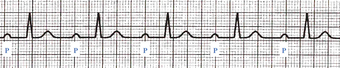
Generally, first-degree AV block is a benign asymptomatic condition that does not require treatment. However, it can indicate disease in the AV node associated with susceptibility to higher degrees of AV block, especially under the effect of drugs that can further impair AV conduction.
Second-Degree AV Block
Second-degree AV block is characterized by intermittent failure of AV conduction, resulting in some P waves that are not followed by a QRS complex.
There are two forms of second-degree AV block. In type I block (also termed Wenckebach block), the degree of AV delay gradually increases with each beat until an impulse is completely blocked (i.e. no QRS after the P wave). The ECG shows a progressive increase in the PR interval from one beat to the next until a single QRS complex is absent, after which the PR interval shortens to its initial length, and the cycle repeats again.
The following ECG tracing shows a second-degree type I AV block. It shows constant P-wave rate, but the PR interval progressively lengthens until a QRS is completely blocked:

Type I block almost always results from impaired conduction in the AV node (rather than more distally in the conduction system). It is usually benign and may be seen in children, athletes, and people with high vagal tone, particularly during sleep. It may also occur during acute myocardial infarction because of increased vagal tone or ischemia of the AV node,.
Treatment of type I block is typically not necessary, but in symptomatic cases, administration of IV atropine or isoproterenol usually improves AV conduction transiently. Occasionally, a permanent pacemaker is required for symptomatic block that does not resolve spontaneously or persist despite the correction of aggravating factors.
Type II second-degree AV block is a more dangerous condition characterized by the sudden intermittent loss of AV conduction, without preceding gradual lengthening of the PR interval. The block may persist for two or more beats (i.e. two sequential P waves not followed by QRS complexes), in which case it is known as high-grade AV block. Type II block is usually caused by conduction block distal to the AV node (in the bundle of His or the Purkinje system), and the QRS pattern often is widened in a pattern of bundle branch block. This type of block may arise from extensive myocardial infarction involving the septum or from chronic degeneration of the His–Purkinje system. It usually indicates severe disease and may progress to complete heart block without warning; therefore, a pacemaker is usually warranted, even in asymptomatic patients.
The following tracing shows second-degree type II AV block. A QRS complex is blocked (after the fourth P wave) without gradual lengthening of the preceding PR intervals. While the QRS width in this example is normal, it is often widened in patients with type II block:

The following ECG tracing shows high degree AV block:

Third-Degree AV Block
Third-degree AV block, also termed complete heart block, is present when there is complete failure of conduction between the atria and ventricles. In adults, the most common causes are acute myocardial infarction and chronic degeneration of the conduction pathways with advanced age.
Third-degree AV block electrically disconnects the atria and ventricles; causing loss of any relation between the P waves and QRS complexes; the atria depolarize in response to SA node activity, while a more distal escape rhythm drives the ventricles independently.
Depending on the site of the escape rhythm, the QRS complexes may be of normal width and occur at 40 to 60 bpm (originating from the AV node) or may be widened and occur at slower rates (originating from the His–Purkinje system). Patients frequently experience light-headedness or syncope, and permanent pacemaker implantation is almost always required.
Note
Third degree AV block is an example of what is called “AV dissociation”, which is a general description for any situation that causes the atria and ventricles to beat independently, without any direct relationship between P waves and QRS complexes.
The following ECG tracing shows third degree AV block. The P wave and QRS rhythms are independent from one another. The QRS complexes are widened as they originate from the distal ventricular conduction system, not at the bundle of His. The second and fourth P waves are superimposed on normal T waves.

Supraventricular Arrhythmias
Sinus Tachycardia
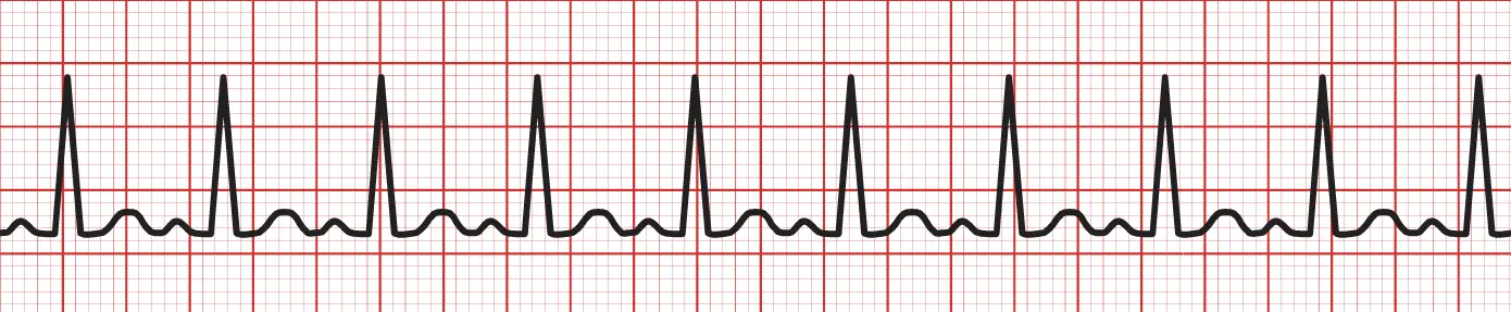
Sinus tachycardia is characterized by an SA node discharge rate greater than 100 bpm with normal P waves and QRS complexes. It most often results from increased sympathetic and/or decreased vagal tone, which is a normal physiological response to exercise.
However, it may also result from pathologic conditions, including fever, hypoxemia, hyperthyroidism, hypovolemia, and anemia. In such cases, sinus tachycardia is usually a sign of the severity of the primary disease, and treatment should be directed at the underlying cause.
The following ECG trace shows sinus tachycardia:
Atrial Premature Beats
Atrial premature beats (APBs) are common in healthy as well as diseased hearts. They originate from automaticity or reentry in an atrial focus outside the SA node and are often exacerbated by sympathetic stimulation. APBs are usually asymptomatic but may cause palpitations.
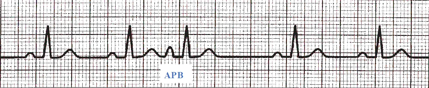
On ECG, an APB appears as an earlier-than-expected P wave with an abnormal shape (i.e. abnormal sequence of conduction through the atria). The QRS complex that follows is usually normal, resembling the QRS during sinus rhythm, because ventricular conduction is not impaired. However, if the abnormal atrial focus fires soon after the previous beat, the impulse may encounter an AV node functional block (i.e. still refractory to excitation), and the premature P wave is not followed by a QRS complex (termed blocked APB).
Similarly, if the ectopic focus fires just a bit later in diastole, it may conduct through the AV node but encounter portions of the His–Purkinje system that are still refractory, and the impulse is conducted through the ventricles more slowly than normal, producing abnormally wide QRS complexes (termed APB with aberrant conduction).
APBs require treatment only if they are symptomatic. Because caffeine, alcohol, and adrenergic stimulation can all predispose to APBs, it is important to address these factors. β-Blockers are the initial preferred pharmacologic treatment if needed.
The following ECG trace shows an atrial premature beat, with a P wave occurring earlier than expected and having an abnormal shape followed by a sinus pause.
Atrial Flutter
Atrial flutter is characterized by rapid, regular atrial activity at a rate of 180 to 350 bpm. Many of these fast impulses reach the AV node during its refractory period and do not conduct to the ventricles, resulting in a slower ventricular rate. Thus, if the atrial rate is 300 bpm and 2:1 block occurs at the AV node, the ventricular rate is 150 bpm.
Because vagal maneuvers (e.g. carotid sinus massage) decrease AV nodal conduction, they increase the degree of block, temporarily slowing the ventricular rate, which allows better visualization of the underlying atrial activity.
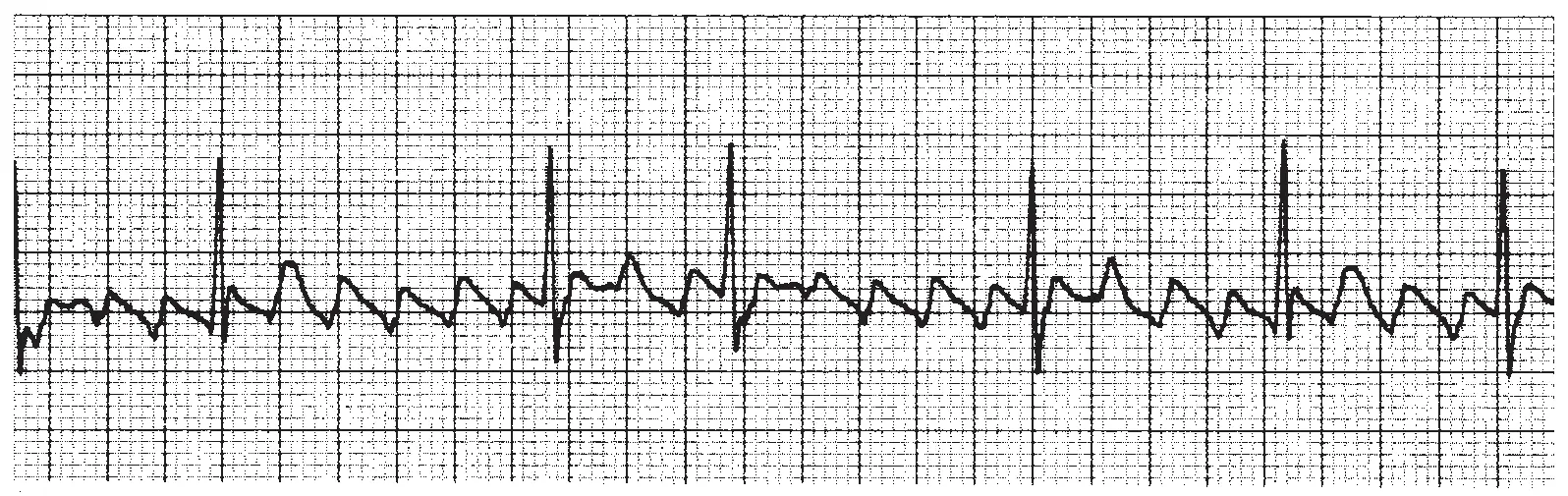
In general, atrial flutter is caused by reentry over a large anatomically fixed circuit. Most commonly, this circuit is the atrial tissue along the tricuspid valve annulus: the circulating wave propagates up the interatrial septum, across the roof and down the free wall of the right atrium, and finally along the floor between the tricuspid valve annulus and inferior vena cava. Because large parts of the atrium are depolarized throughout the cycle, P waves often have a “sawtooth” appearance.
The following ECG trace shows atrial flutter with rapid “saw-toothed” atrial activity:
Atrial flutter generally occurs in patients with preexisting heart disease. It may be paroxysmal (lasting for less than a week), persistent (lasting for weeks), or permanent. Symptoms of atrial flutter depend on the accompanying ventricular rate. If the rate is less than 100 bpm, the patient may be asymptomatic. Faster rates often cause palpitations, dyspnea, or weakness.
Paradoxically, antiarrhythmic medications that reduce the rate of atrial flutter may make the rhythm more dangerous by allowing the AV node more time to recover between impulses. For example, a patient with atrial flutter at a rate of 280 bpm and 2:1 conduction at the AV node would have a ventricular rate of 140 bpm. If the atrial rate then slows to 220 bpm, the AV node may be able to recover sufficiently between depolarizations to conduct every atrial impulse, causing the ventricular rate to accelerate to 220 bpm.
Atrial flutter also predisposes to atrial thrombus formation, and anticoagulation therapy is often appropriate. Several approaches for the conversion of atrial flutter to sinus rhythm are available:
-
For symptomatic patients with atrial flutter (either acute or chronic), the most expeditious therapy is cardioversion to restore sinus rhythm.
-
Flutter can be terminated by burst pacing using a temporary or permanent pacemaker.
-
Patients without an immediate need for cardioversion can begin pharmacologic therapy. First, the ventricular rate is slowed by drugs that increase AV block: β-blockers, certain calcium channel blockers (e.g. verapamil, diltiazem), or digoxin. Once ventricular rate is effectively slowed, attempts can be made to restore sinus rhythm using antiarrhythmic drugs (usually class IC or class III agents). Should these drugs fail to convert the rhythm, electrical cardioversion can be undertaken. Once sinus rhythm has been restored, antiarrhythmic drugs may be administered chronically to prevent recurrence.
-
For chronic therapy to prevent recurrences, catheter ablation is often a better alternative than pharmacologic approaches. In this method, an electrode catheter is inserted into the femoral vein, passed via the inferior vena cava to the right atrium, and used to localize and cauterize part of the reentrant loop to permanently interrupt the flutter circuit.
Atrial Fibrillation
AF is a chaotic rhythm with an atrial rate so fast (350 to 600 discharges/min) that distinct P waves are not discernible on the ECG. As with atrial flutter, many of the atrial impulses encounter refractory blocks at the AV node, allowing only some of the depolarizations to be conducted to the ventricles in a very irregular fashion (indicated by a characteristic “irregularly irregular” rhythm).
The average ventricular rate in untreated AF is approximately 140 to 160 bpm. Because discrete P waves are not visible on the ECG, the baseline shows low-amplitude undulations punctuated by QRS complexes and T waves.

The mechanism of AF likely involves multiple reentrant circuits within the atria, and in some patients, the rhythm repetitively shifts between fibrillation and flutter. To sustain AF, a minimum number of reentrant circuits is needed, and an enlarged atrium increases the potential for this to occur. Thus, AF is often associated with atrial enlargement. Accordingly, diseases that increase atrial pressure and size promote AF, including heart failure, hypertension, coronary artery disease, pulmonary disease, and cardiomyopathies. Thyrotoxicosis and alcohol consumption can also precipitate AF in some individuals. In addition, AF is a common rhythm disturbance in the elderly.
Note
If AF is caused by alcoholism, it is sometimes referred to as Holiday heart syndrome
As with atrial flutter, when the ventricular rate is less than 100 beats/min, AF may be asymptomatic. Faster ventricular rates may compromise cardiac output, resulting in hypotension and pulmonary congestion (especially in patients with a stiffened left ventricle in whom the loss of normal atrial contraction can significantly reduce left ventricular filling and stroke volume). AF is also an important cause of stroke. The absence of organized atrial contraction promotes blood stasis in the atria, increasing the risk of thrombus formation in the left atrial appendage, which can embolize to the cerebral circulation and other systemic sites.
Thus, treatment of AF is directed at three aspects of the arrhythmia: (1) ventricular rate control, (2) assessment of the need for anticoagulation, and (3) consideration of methods to restore sinus rhythm.
Antiarrhythmic drug treatment of AF is similar to that of atrial flutter. β-Blockers or certain channel antagonists (diltiazem, verapamil) are administered to promote block at the AV node, reducing ventricular rate. Digitalis is less effective in AF, although it may be useful in patients with accompanying impairment of ventricular contractile function.
For those who remain symptomatic despite adequate rate control, cardioversion is usually attempted either chemically (using class IC, IA, or III antiarrhythmic drugs) or electrically. Following successful conversion to sinus rhythm, antiarrhythmic drugs are often continued to prevent recurrences. However, because of the potential side effects, in patients with asymptomatic AF, it is often appropriate to simply control the ventricular rate and give anticoagulation therapy chronically, rather than to pursue cardioversion. Such approach is supported by clinical trials of AF that have assessed long-term clinical outcomes.
Note
Remember that antiarrhythmic therapy alone can paradoxically increase the ventricular rate, and so ventricular rate control through β-Blockers must be attempted first before any antiarrhythmic drugs are given.
AF persistent for hours may predispose to atrial thrombus formation, and systemic anticoagulation for at least 3 weeks is usually warranted prior to cardioversion. Alternatively, a transesophageal echocardiogram can be performed to evaluate for the presence of thrombus; if none is found, cardioversion may proceed directly, provided that anticoagulation is instituted for several weeks thereafter (since recovery of normal atrial contraction may be delayed for a period of time following cardioversion).
Because the efficacies and toxicities of antiarrhythmic drugs have been disappointing, nonpharmacologic options for the management of AF have been devised. For example, the surgical maze procedure places multiple incisions in the left and right atria to prevent the formation of reentry circuits and is sometimes performed in patients undergoing cardiac surgery for coronary artery or valve disease who also have AF. A less invasive approach is percutaneous catheter ablation, however, this requires extensive catheter manipulation and ablation in the left atrium, and risks includes stroke from systemic thromboembolism or cardiac perforation that can cause pericardial tamponade. Thus, catheter ablation for AF is usually reserved for patients who remain symptomatic despite pharmacologic approaches.
When sinus rhythm cannot be maintained and the heart rate cannot be controlled adequately with medications, catheter ablation of the AV node is another available procedure. This method intentionally creates complete heart block as a means to permanently slow the ventricular rate. Permanent ventricular pacemaker placement is then also required to generate an adequate ventricular rate.
Focal Atrial Tachycardia
Focal atrial tachycardia (AT) results from either automaticity of an atrial ectopic site or reentry. The ECG has the appearance of sinus tachycardia, with a P wave before every QRS complex, but the P-wave morphology is different from that of sinus rhythm, indicating depolarization of the atrium from an abnormal location.
The arrhythmia can be paroxysmal and of limited duration, or it can persist. Short, asymptomatic bursts of AT are commonly observed on 24-hour ECG recordings, even in healthy people.
AT can be caused by digitalis toxicity and is also aggravated by elevated sympathetic tone. Initial treatment includes correction of any contributing factors. Unlike AV related atrial tachycardia, vagal maneuvers (such as carotid sinus massage) may have no effect on discharges from an ectopic focus. However, β-blockers; calcium channel blockers; and class IC, IA, and III antiarrhythmic drugs can be effective. Catheter ablation is also a useful option for symptomatic patients.
Multifocal Atrial Tachycardia
In multifocal atrial tachycardia (MAT), the ECG shows an irregular rhythm with at least 3 different P-wave morphologies, and the average atrial rate is greater than 100 bpm:
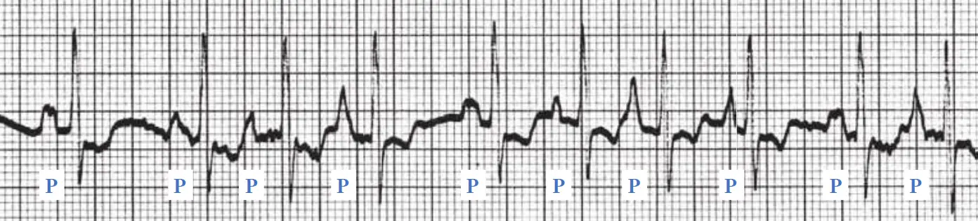
An isoelectric (i.e. “flat”) baseline between P waves distinguishes MAT from the chaotic baseline of AF. This rhythm is likely caused by either abnormal automaticity in several foci within the atria or triggered activity. It occurs most often in the setting of severe obstructive pulmonary disease and hypoxemia.
Because patients with this rhythm are often critically ill from the underlying disease, the mortality rate is high, and treatment is aimed at the causative disorder. The calcium channel blocker verapamil is often effective at slowing the ventricular rate as a temporizing measure.
Paroxysmal Supraventricular Tachycardia
Paroxysmal supraventricular tachycardias (PSVTs) manifest with (1) sudden onset and sudden termination, (2) atrial rates between 140 and 250 bpm, and (3) narrow (normal) QRS complexes, unless aberrant conduction is present. The mechanism of PSVTs is most often reentry involving the AV node, atrium, or an accessory pathway between an atrium and a ventricle. Enhanced automaticity and triggered activity in the atrium or AV node are less common causes.
AV Nodal Reentrant Tachycardia (AVNRT)
AVNRT is the most common form of PSVT. In the normal heart, the AV node consists of a compact portion and several atrial extensions. The latter constitute two or more potential pathways for conduction through the AV node. In some people, these extensions have different conduction times, providing both slow- and fast-conducting pathways.
The fast pathway is characterized by a rapid conduction velocity, whereas the slow pathway demonstrates slower conduction but typically has a shorter refractory period. Thus, although the fast pathway conducts rapidly, it takes longer to recover between impulses compared with the slow pathway. Normally, a stimulus arriving at the AV node travels down both pathways, but the impulse traveling down the fast pathway reaches the bundle of His first. By the time the impulse traversing the slow pathway reaches the bundle of His, it encounters refractory tissue and is blocked. Thus, under normal conditions, only the fast pathway impulse makes its way to the ventricles.
In contrast, consider what happens when an APB spontaneously occurs; because the refractory period of the fast pathway is relatively long, an APB would find that pathway unexcitable. However, the impulse is able to conduct over the slow pathway (which is excitable because of its shorter refractory period). By the time this impulse travels down the slow pathway, the distal end of the fast pathway may have had time to repolarize, and the impulse is able to propagate both distally (to the bundle of His) and backward to the atria, up the fast pathway in a retrograde direction. On reaching the atria, the impulse can then circulate back down the slow pathway, completing the reentrant loop.
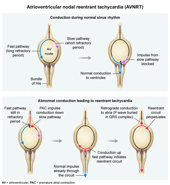
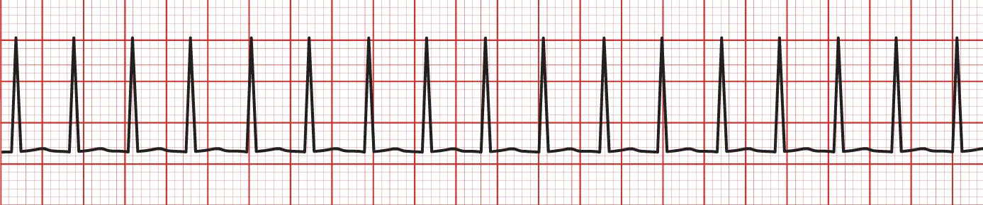
ECG in AVNRT shows a regular tachycardia with normal-width QRS complexes. P waves may not be apparent, because retrograde atrial depolarization typically occurs simultaneously with ventricular depolarization (P waves may be “buried” in QRS complexes). When P waves are visible, they are superimposed on the terminal portion of the QRS complex and inverted (negative deflection) in limb leads II, III, and aVF, because of the craniocaudal direction of atrial activation.
The following ECG trace shows PSVT with P waves “buried” in the QRS complexes:
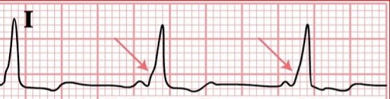
Rarely, the reentrant loop revolves in the reverse direction, with anterograde conduction down the fast pathway and retrograde conduction up the slow pathway. This is known as uncommon AVNRT and, unlike the more common rhythm, typically results in clearly visible retrograde P waves following the QRS complex on the ECG.
AVNRT often presents in teenagers or young adults. It is usually well tolerated but causes palpitations that many find frightening, and rapid tachycardias can cause lightheadedness or shortness of breath. In elderly patients or those with underlying heart disease, more severe symptoms may result, such as syncope, angina, or pulmonary edema.
Acute treatment of AVNRT is aimed at terminating reentry by impairing conduction in the AV node. Valsalva maneuver or carotid sinus massage may increase the vagal tone and stop the tachycardia. The most rapidly effective pharmacologic treatment is IV adenosine. Other drug options include IV calcium channel antagonists (verapamil and diltiazem) or β-blockers. Most patients have infrequent episodes that terminate with vagal maneuvers and do not require pharmacologic interventions. Frequent symptomatic episodes warrant preventive therapy using oral β-blockers, calcium channel blockers, or digoxin. Catheter ablation of the slow AV nodal pathway is usually curative and recommended when pharmacologic therapy fails or is not desired by the patient, but has a small risk () of heart block owing to unintended damage to the fast AV nodal pathway, a complication that requires permanent pacemaker implantation.
Atrioventricular Reentrant Tachycardias
Atrioventricular reentrant tachycardias (AVRTs) are similar to AVNRTs except that in the former, one limb of the reentrant loop is constituted by an accessory pathway (bypass tract), rather than by separate fast and slow pathways in the AV node itself.
Approximately 1 in 1,500 people has such a pathway congenitally. Accessory pathways allow an impulse to conduct from atrium to ventricle (anterograde conduction), from ventricle to atrium (retrograde conduction), or in both directions. Depending on the characteristics of the pathway, one of two characteristic entities can result: (1) ventricular preexcitation syndrome or (2) PSVT resulting from a concealed accessory pathway. Some pathways do not conduct impulses at rates sufficient to cause tachycardias and cause no symptoms at all.
Ventricular Preexcitation Syndrome
In patients with ventricular preexcitation (also termed Wolff–Parkinson–White [WPW] syndrome), atrial impulses can pass to the ventricles through both the AV node and the accessory pathway. Because conduction through the accessory pathway is usually faster than that via the AV node, the ventricles are stimulated earlier than by normal conduction through the AV node. This premature ventricular stimulation from the accessory pathway causes a characteristic ECG appearance:
-
Shortened PR interval ( seconds) because ventricular stimulation begins earlier than normal through the accessory pathway
-
The QRS has a slurred upstroke (referred to as a delta wave) because the initial ventricular activation by the accessory pathway is slower than activation over the Purkinje system
-
The QRS complex is widened because it represents fusion of two excitation waves through the ventricles, one from the accessory pathway and one from the normal His–Purkinje system
Patients with WPW syndrome are predisposed to PSVTs because the accessory pathway provides a potential limb for a reentrant loop. The most common PSVT in these patients is orthodromic AVRT, where an impulse travels anterogradely down the AV node to the ventricles and then retrogradely up the accessory tract to the atria. Because the ventricles in this situation are depolarized exclusively via the normal conduction system, there is no delta wave during the tachycardia and the width of the QRS is normal. Retrograde P waves are often visible soon after each QRS complex because the atria are stimulated from below via retrograde conduction through the accessory pathway.
In fewer than 10% of patients with AVRT involving an accessory pathway, the reentrant arrhythmia travels in the opposite direction; impulses travel anterogradely down the accessory pathway and retrogradely up the AV node. Termed antidromic AVRT, its ECG is characterized by a wide QRS complex because the ventricles are activated entirely from anterograde conduction over the accessory pathway. From ECG alone, such antidromic tachycardia is difficult to distinguish from ventricular tachycardia.
A third type of arrhythmia encountered in patients with WPW syndrome is anterograde conduction over the accessory pathway when AF or atrial flutter is present. Some accessory pathways have short refractory periods that allow faster rates of ventricular stimulation than the AV node. Thus, during AF or atrial flutter, ventricular rates as fast as 300 bpm may be seen. Such rates are poorly tolerated and can lead to ventricular fibrillation and cardiac arrest, even in a young, otherwise healthy patient.
Pharmacologic management of arrhythmias in patients with WPW syndrome requires greater caution than that of AVNRTs. Although digitalis, β-blockers, and certain calcium channel blockers are effective at blocking conduction through the AV node, they do not slow conduction over most accessory pathways. Sometimes these drugs actually shorten the refractory period of the accessory pathway, thus speeding conduction. In contrast, sodium channel blockers (specifically, class IA and IC antiarrhythmics) and some class III antiarrhythmic drugs slow conduction and prolong the refractory period of accessory pathways as well as the AV node; therefore, these are the preferred pharmacologic agents for this condition.
When a patient with WPW presents with a tachycardia, acute therapy depends on the patient’s tolerance of the arrhythmia. If accompanied by hemodynamic collapse, immediate cardioversion is required. Conversely, if the patient is hemodynamically stable, IV procainamide (a class IA agent that slows conduction in the accessory pathway) or ibutilide (a class III agent that prolongs refractoriness in the accessory pathway) will often terminate the arrhythmia.
Patients who have WPW with symptomatic arrhythmias should generally undergo invasive radiofrequency ablation of the accessory pathway. If this procedure is not an option, chronic oral therapy should include a drug that slows accessory pathway conduction (i.e. class IA, IC, or III agent).
The Lown–Ganong–Levine syndrome is also characterized by a short PR interval but a normal, narrow QRS complex (i.e. no delta wave). It used to be considered a form of preexcitation, but most patients just have enhanced conduction through the normal AV node, thus shortening the PR interval. When PSVT occurs in these patients, it is usually simply due to AV nodal reentry.
Concealed Accessory Pathways
Accessory pathways do not always result in ECG findings of ventricular preexcitation. Many are capable of only retrograde conduction. In this case, during sinus rhythm, the ventricles are depolarized normally through the AV node alone and the ECG is normal. However, because the accessory pathway is capable of retrograde conduction, it can form a limb of a reentrant circuit under appropriate circumstances and result in orthodromic AVRT.
Management of patients with tachycardia involving a concealed accessory pathway is the same as for patients with AVNRT. Because the reentrant circuit travels anterogradely down the AV node, vagal maneuvers and drugs that interrupt conduction over the AV node can terminate the tachycardia. Another option for recurrent episodes is catheter ablation of the accessory pathway, which is curative in most patients.
Ventricular arrhythmias
Ventricular arrhythmias include ventricular premature beats, ventricular tachycardia, and ventricular fibrillation. Ventricular arrhythmias are usually more dangerous than supraventricular rhythm disorders and are responsible for many of the sudden cardiac deaths that occur every year in the US.
Ventricular Premature Beats (VPBs)
A VPB arises when an ectopic ventricular focus fires. On ECG, a VPB appears as a widened QRS complex, because the impulse travels from its ectopic site through the ventricles via slow cell-to-cell connections rather than through the normal rapidly conducting His–Purkinje system. Furthermore, the ectopic beat is not related to a preceding P wave. VPBs are usually followed by a long baseline period known as a compensatory pause as the surrounding conduction tissue and the AV node enter a refractory period from the premature ventricular excitation.
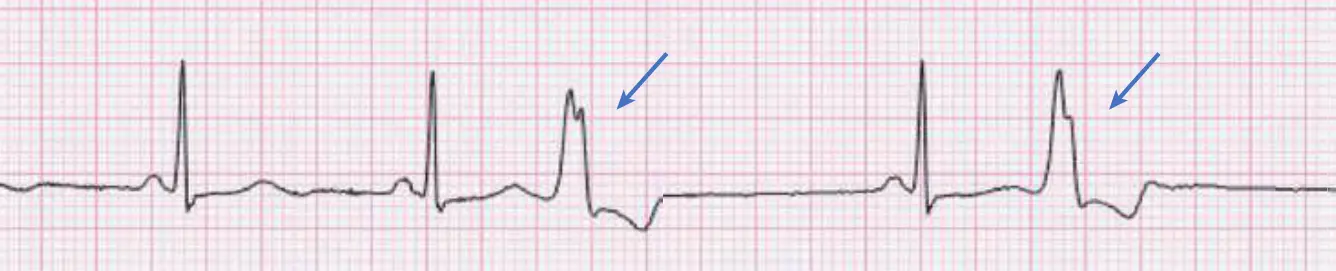
When every alternate beat is a VPB, the rhythm is termed bigeminy. When two normal beats precede every VPB, it is termed trigeminy. Consecutive VPBs are referred to as couplets or triplets.
Similar to APBs, VPBs are common even among healthy people and are often asymptomatic and benign. Specific precipitants of VPBs include medications (e.g. β-adrenergic agonists), caffeine, electrolyte abnormalities (e.g. hypokalemia, hypomagnesemia), and hypoxia.
In patients without heart disease, they confer no added risk of a life-threatening arrhythmia. They can, however, be an indication of an underlying cardiac disorder. They also have been associated with an increased risk of sudden death in patients with heart failure or prior myocardial infarction.
In otherwise healthy persons, treatment of VPBs mainly involves reassurance and, if needed, symptomatic control using β-blockers. In patients with advanced heart disease with features that place them at risk of life-threatening arrhythmias, placement of an ICD is typically recommended.
Ventricular Tachycardia
VT is a sequence of 3 or more consecutive VPBs. If it persists for more than 30 seconds, produces severe symptoms, or requires termination by cardioversion or administration of an antiarrhythmic drug, it is designated as sustained VT; shorter, self-terminating episodes are termed non-sustained VT.
Both forms are found most commonly in patients with structural heart disease, including myocardial ischemia and infarction, heart failure, ventricular hypertrophy, primary electrical diseases (e.g. long-QT syndromes), valvular heart diseases, and congenital cardiac abnormalities.
The QRS complexes of VT are typically wide ( seconds) and occur at a rate of 100 to 200 bpm or sometimes faster. VT is further categorized according to its QRS morphology. When every QRS complex appears the same and the rate is regular, it is referred to as monomorphic VT. Sustained monomorphic VT usually indicate a structural abnormality that supports a reentry circuit, most commonly a region of myocardial fibrosis. Occasionally, sustained monomorphic VT occurs as a result of an ectopic ventricular focus in an otherwise healthy person (referred to as idiopathic VT).
When the QRS complexes continually change in shape and the rate varies from beat to beat, it is referred to as polymorphic VT. Multiple ectopic foci or a continually changing reentry circuit is the cause. Torsade de pointes and acute myocardial ischemia or infarction are the most common causes of polymorphic VT.
The symptoms of VT vary depending on the rate, duration, and underlying condition of the heart. Sustained VT can cause low cardiac output resulting in syncope, pulmonary edema, or progress to cardiac arrest. These consequences are most likely in patients with an underlying depressed contractile function. Conversely, if sustained VT is at a relatively slow rate (e.g. bpm), it may be well tolerated and cause only palpitations.
Sustained episodes of VT are dangerous because they can produce syncope or deteriorate into VF, which is fatal if not quickly corrected. Acute treatment usually consists of electrical cardioversion. Intravenous administration of certain antiarrhythmic drugs, such as amiodarone, procainamide, or lidocaine, can be considered if the patient is hemodynamically stable. After sinus rhythm is restored, careful evaluation is required to define whether underlying structural heart disease is present and to correct any aggravating factors, such as myocardial ischemia, electrolyte disturbances, or drug toxicities. Patients who had VT in the setting of structural heart disease have a high risk of recurrence and sudden cardiac death; and implantation of an ICD is usually warranted.
Patients who experience VT in the absence of underlying structural heart disease are usually found to have idiopathic VT, which is rarely life threatening; β-Blockers, calcium channel blockers, or catheter ablation is commonly effective.
Genetic Mutations and Ventricular Arrhythmias
Examples of inherited structural disease that can be complicated by life-threatening ventricular arrhythmias include hypertrophic cardiomyopathy, familial dilated cardiomyopathies, and arrhythmogenic right ventricular cardiomyopathy (ARVC) described in part 9.
ARVC may be suspected on ECG by the presence of inverted T waves in leads V1 through V3 and occasionally an epsilon wave (terminal notch of the QRS complex) in lead V1, which reflects abnormal RV activation.
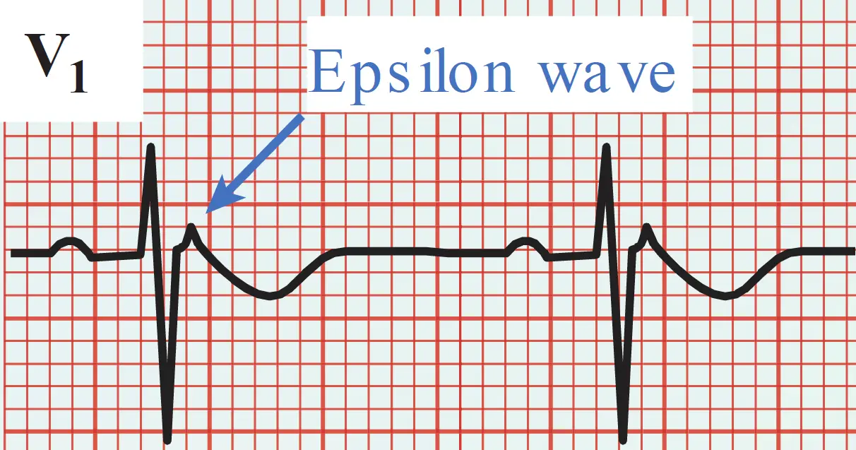
Several other inherited arrhythmic disorders occur in the absence of structural cardiac disease. These occur infrequently but are important because they can cause life-threatening polymorphic VT or VF in young, otherwise healthy people without prior warning. The most common of these conditions are (1) Brugada syndrome, (2) congenital long QT syndrome, and (3) familial catecholaminergic polymorphic VT.
Brugada syndrome is an autosomal dominant disease and has been linked in some (but not all) families to mutations in a sodium channel subunit gene (SCN5A). A clue to the presence of this syndrome is a specific ECG finding of prominent ST elevation in leads V1 through V3. This pattern may be present chronically or intermittently; in the latter case, the syndrome may be unmasked by administering sodium channel blocking antiarrhythmic drugs.
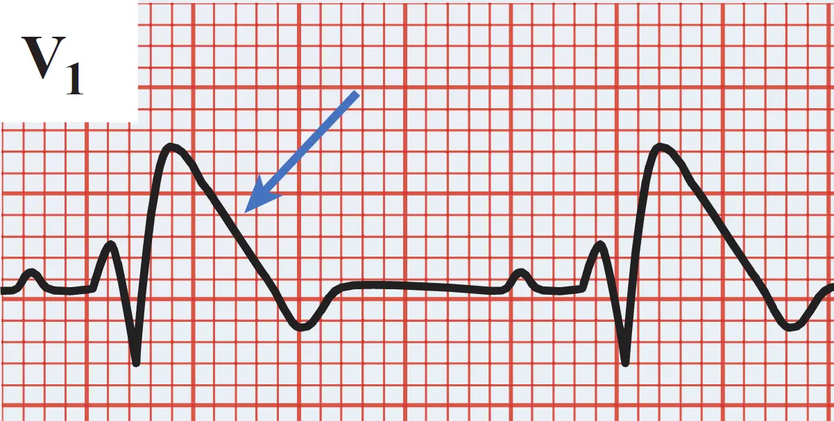
Congenital long QT syndromes are associated with prolonged ventricular repolarization (hence the long QT interval), which can lead to life-threatening polymorphic VT (i.e. Torsade de pointes). Mutations in a number of different genes can cause this syndrome by prolonging the action potential duration. Most identified mutations alter ion channel function to either enhance the depolarizing Na+ current or impair the repolarizing K+ current.
Inheritance pattern, gene penetrance, and symptomatology of patients with long QT syndrome is highly variable, even for individuals with the same mutation. The degree of QT prolongation and, in some cases, patient’s gender are predictors of arrhythmic risk. An affected patient may be asymptomatic and come to medical attention only as a result of the abnormal ECG or because of a family member who died suddenly. Others present with syncope or sudden death due to Torsade de pointes. The most common forms (LQT1 and LQT2) are associated with ventricular arrhythmias during physical exercise (particularly swimming) or emotional stress. Conversely, those with LQT3 are much more likely to experience cardiac arrest at rest or during sleep.
Other acquired conditions that further prolong the QT interval can trigger life-threatening arrhythmias in patients with long QT syndrome, including hypokalemia, hypomagnesemia, hypocalcemia, and several medications (including many antiarrhythmic drugs). Conversely, β-blockers reduce the risk of arrhythmias in many forms of congenital LQTS, even though they do not shorten the QT interval.
Familial catecholaminergic polymorphic VT, inherited in autosomal dominant and recessive patterns, is marked by VT and/ or VF during exercise or emotional arousal. The mechanism is thought to be triggered activity resulting from delayed afterdepolarizations. Mutations in affected families have been demonstrated in at least two genes involved in intracellular calcium handling, including a missense mutation in the gene for the cardiac ryanodine receptor. Treatment with a β-blocker, calcium channel blocker, and/ or drug that inhibits ryanodine receptor–mediated calcium release (e.g. the class IC antiarrhythmic flecainide has this additional attribute) may be effective.
Note
For patients with any of these genetic syndromes that are at high risk of life-threatening arrhythmias, ICD implantation is warranted.
Distinguishing Monomorphic VT from Supraventricular Tachycardia
VT can usually be distinguished from SVT by the width of the QRS complex: it is routinely wide in the former and narrow (i.e. normal) in the latter. However, under certain circumstances, SVT can result in wide QRS complexes and may appear similar to monomorphic VT. This is termed SVT with aberrant ventricular conduction, or simply SVT with aberrancy, and may arise due to:
-
A patient has an underlying conduction abnormality (e.g. bundle branch block), such that the QRS is wide even in normal sinus rhythm
-
A patient develops antidromic tachycardia through an accessory pathway.
Certain features can help distinguish wide QRS complexes of monomorphic VT from those of supraventricular rhythms with aberrant conduction. In patients with a history of prior myocardial infarction, congestive heart failure, or left ventricular dysfunction, a wide complex tachycardia is more likely to be VT rather than SVT with aberrancy. At the bedside, SVT is more probable if vagal maneuvers (e.g. carotid sinus massage) affect the rhythm.
Electrocardiographically, SVT is more likely if the morphology of the QRS at the rapid rate is similar to that on the patient’s ECG tracing obtained while in sinus rhythm. Conversely, VT is more likely if there is no relationship between the QRS complexes and any observed P waves (AV dissociation) or the QRS complexes in each of the chest leads (V1 through V6) have a similar appearance, with a dominant positive or negative deflection (i.e. there is “concordance” of precordial QRS complexes).
Other morphologic ECG features have been used to distinguish VT from SVT with aberrancy, but the distinction is often very difficult. Most patients with wide QRS tachycardia should be managed as though they have VT until proven otherwise.
Torsade de Pointes
Torsade de pointes (“twisting of the points”) is a form of polymorphic VT that presents as varying amplitudes of the QRS, as if the complexes were “twisting” about the baseline. It can result from early afterdepolarizations, particularly in patients who have a prolonged QT interval.
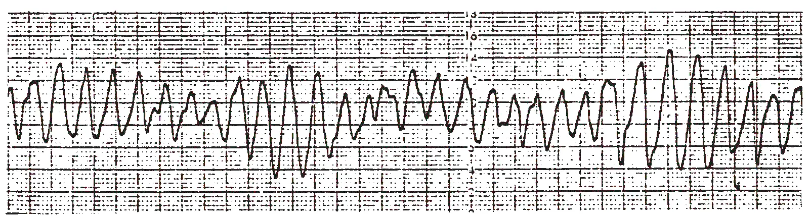
QT prolongation can result from:
-
Electrolyte disturbances particularly hypokalemia or hypomagnesemia.
-
Drugs that block cardiac potassium channels including many antiarrhythmic drugs (particularly class III drugs and some class I drugs).
-
Many non-cardiac medications, including erythromycin, phenothiazines, haloperidol, and methadone.
-
Hereditary ion channel abnormalities (like the ones we talked about above).
Torsade de pointes is usually symptomatic, causing light-headedness or syncope, but is frequently self-limited. Its main danger results from degeneration into VF.
When it is drug or electrolyte induced, correcting the underlying cause resolves the episodes. However, measures to suppress episodes are usually required to allow time to address the cause. These include IV magnesium, and pharmacological therapy to shorten the QT interval and increase the heart rate like IV β-adrenergic stimulating agents (e.g. isoproterenol).
When torsade de pointes results from congenital prolongation of the QT interval, β-Blockers are the treatment of choice, because sympathetic stimulation actually aggravates the arrhythmia
Ventricular Fibrillation
VF is an immediately life-threatening arrhythmia. It results in disordered, rapid stimulation of the ventricles with no coordinated contractions. The result is essentially cessation of cardiac output and death if not quickly reversed. This rhythm most often occurs in patients with severe underlying heart disease and is the major cause of mortality in acute myocardial infarction.
VF is often initiated by an episode of VT, which degenerates, it is believed, by the breakup of excitation waves into multiple smaller wavelets of reentry that wander through the myocardium.
On the ECG, VF is characterized by a chaotic irregular appearance without discrete QRS complexes:
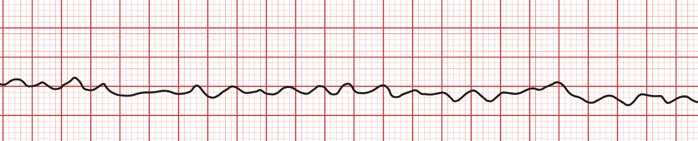
Untreated, VF rapidly leads to death. The only effective therapy is prompt electrical defibrillation. As soon as the heart has been converted to a safe rhythm, the underlying precipitant of the arrhythmia (e.g. myocardial ischemia, electrolyte imbalances, hypoxemia, or acidosis) should be corrected to prevent further episodes. IV antiarrhythmic drugs may be administered to prevent immediate recurrences. If no reversible inciting precipitant is found, survivors of VF usually receive an ICD.
See also
References
- Mechanisms of cardiac arrhythmias. (2016). In L. S. Lilly (Ed.), Pathophysiology of heart disease: A collaborative project of medical students and faculty (6th ed., pp. 268–286). Wolters Kluwer.
- Clinical aspects of cardiac arrhythmias. (2016). In L. S. Lilly (Ed.), Pathophysiology of heart disease: A collaborative project of medical students and faculty (6th ed., pp. 287–309). Wolters Kluwer.
Additional Reading
- Chen PS, Joung B, Shinohara T, et al. The initiation of the heart beat. Circ J. 2010;74:221–225.
- DiFrancesco D. The role of the funny current in pacemaker activity. Circ Res. 2010;106:434–446
- Grant AO. Cardiac ion channels. Circ Arrhythm Electrophysiol. 2009;2:185–194.
- Katritsis DG, Camm AJ. Atrioventricular nodal reentrant tachycardia. Circulation. 2010;122:831–840.
- Spector P. Principles of cardiac electric propagation and their implications for re-entrant arrhythmias. Circ Arrhythm Electrophysiol. 2013;6:655–661
- January CT, Wann LS, Alpert JS, et al. 2014 AHA/ ACC/ HRS guideline for the management of patients with atrial fibrillation: A report of the American College of Cardiology/American Heart Association Task Force on Practice Guidelines and the Heart Rhythm Society. Circulation. 2014;130(23):2071–2104.
- Link MS. Clinical practice. Evaluation and initial treatment of supraventricular tachycardia. N Engl J Med. 2012;367:1438–1448
- Pediatric Congenital Electrophysiology Society, Heart Rhythm Society, American College of Cardiology Foundation, et al. PACES/ HRS expert consensus statement on the management of the asymptomatic young patient with a Wolff-Parkinson-White (WPW, ventricular preexcitation)electrocardiographic pattern. Heart Rhythm. 2012;9:1006–1024.
- Roden DM. Clinical practice. Long-QT syndrome. N Engl J Med. 2008;358:169–176.
- Stevenson WG. Current treatment of ventricular arrhythmias: State of the art. Heart Rhythm. 2013;10:1919–1926.
- Tracy CM, Epstein AE, Darbar D, et al. 2012 ACCF/ AHA/ HRS focused update of the 2008 guidelines for device-based therapy of cardiac rhythm abnormalities: A report of the American College of Cardiology Foundation/ American Heart Association Task Force on Practice Guidelines. J Am Coll Cardiol. 2012;60:1297–1313