Part 9: Cardiomyopathies
Wed Jul 29 2026
By B. Hassan

Cardiomyopathies are a diverse set of heart muscle disorders that cause dysfunction of the myocardium. Excluded from the definition of this group of diseases is heart muscle impairment due to other specific cardiovascular disorders such as hypertension, valvular abnormalities, or congenital heart disease. Cardiomyopathies often result in inappropriate ventricular hypertrophy or dilatation, and progressive heart failure. These conditions can involve the heart alone or may be a component of a systemic syndrome.
Cardiomyopathies can be classified into 3 main types: dilated cardiomyopathy (DCM), characterized by ventricular chamber enlargement with impaired systolic contractile function; hypertrophic cardiomyopathy (HCM), characterized by an abnormally thickened ventricular wall with abnormal diastolic relaxation but usually intact systolic function; and restrictive cardiomyopathy, characterized by an abnormally stiffened myocardium leading to impaired diastolic relaxation, but systolic contractile function is typically normal or near normal.
Dilated cardiomyopathy
Many different etiologies can result in cardiac enlargement in DCM, including genetic, inflammatory, toxic, and metabolic causes. DCM can also be idiopathic.
Inflammatory causes of DCM include infections (especially viral infections), connective tissue diseases, or peripartum cardiomyopathy.
Toxic causes of DCM include chronic alcoholism and specific chemotherapy agents (i.e. doxorubicin, trastuzumab).
Metabolic causes include hypothyroidism, chronic hypocalcemia, or chronic hypophosphatemia.
Acute viral myocarditis generally affects young, previously healthy people. Common infectious agents include coxsackievirus group B, parvovirus B19, and adenovirus among many others. Viral myocarditis is usually a self-limited disease with full recovery, but for unknown reasons, some patients progress to DCM. It is hypothesized that myocardial destruction and fibrosis result from immune-mediated injury triggered by viral constituents. Nonetheless, immunosuppressive drugs have not been shown to improve the prognosis.
Ventricular biopsy during acute myocarditis may demonstrate active inflammation, but specific viral genomic sequences have been demonstrated in only a minority of patients.
DCM may develop in people with excessive and chronic alcohol consumption. Although the pathogenesis is unknown, ethanol is thought to impair cellular function by impacting mitochondrial oxidative function, myofilament protein synthesis, and cytosolic calcium levels. While its clinical presentation and histologic features are similar to those of other dilated cardiomyopathies, alcoholic cardiomyopathy is important to identify because it is potentially reversible after cessation of ethanol consumption.
Peripartum cardiomyopathy is a form of DCM that presents with heart failure symptoms between the last month of pregnancy and up to 6 months postpartum. Risk factors include advanced maternal age, being African American, and multigravidity (having multiple pregnancies). Ventricular function returns to normal in approximately 50% of affected women following pregnancy, but recurrences of DCM with subsequent pregnancies have been reported.
Note
Out of all the etiologies, the reversible causes of DCM include peripartum cardiomyopathy, alcohol-related DCM, exposure to toxic drugs, and hypothyroidism.
Several familial forms of DCM have been identified and are believed to be responsible for 20% to 30% of what were once classified as idiopathic DCM. Autosomal dominant, autosomal recessive, X-linked, and mitochondrial patterns of inheritance have all been described, leading to defects in contractile force generation, force transmission, energy production, and myocyte viability.
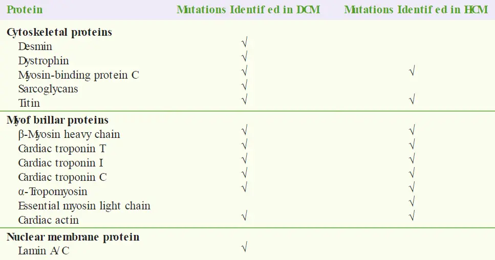
The following table shows some of the identified gene mutations related to cardiomyopathy (including DCM and HCM):
Pathophysiology
The hallmark of DCM is ventricular dilatation with decreased contractile function. Marked enlargement of all four cardiac chambers is typical, though sometimes the disease is limited to the left or right side of the heart. The thickness of the ventricular walls may be increased, but chamber dilatation is out of proportion to any hypertrophy.
Microscopically, there is evidence of myocyte degeneration with irregular hypertrophy and atrophy of myofibers. Interstitial and perivascular fibrosis is often extensive.
As ventricular stroke volume and cardiac output decline because of impaired myocyte contractility, compensatory mechanisms are activated. These include the Frank–Starling mechanism and neurohormonal activation (through the sympathetic and the renin–angiotensin–aldosterone system). As described in part 8, these compensatory mechanisms may render the patient asymptomatic during the early stages of the disease, but they ultimately prove detrimental, causing progressive ventricular remodeling, myocyte degeneration, volume overload, and symptoms of heart failure.
Note
The clinical manifestations of DCM are those of congestive heart failure, refer to part 8 for clinical presentation of heart failure.
As the ventricles enlarge over time, the mitral and tricuspid valves may fail to close properly in systole, and valvular regurgitation ensues. This regurgitation has 3 detrimental consequences: (1) excessive volume and pressure loads are placed on the atria, causing them to dilate, often leading to atrial fibrillation; (2) regurgitation of blood into the left atrium further decreases forward stroke volume; and (3) when the regurgitant volume returns to the LV during each diastole, an even greater volume load is presented to the dilated LV.
Diagnostic Studies
Chest radiography shows an enlarged cardiac silhouette. If heart failure has developed, then pulmonary vascular redistribution, interstitial and alveolar edema, and pleural effusions are evident.
An ECG usually demonstrates atrial and ventricular enlargement. Patchy fibrosis of the myofibers results in a variety of arrhythmias, most importantly atrial fibrillation and ventricular tachycardia. Conduction defects (bundle branch block) are common. In addition, regions of dense myocardial fibrosis may produce localized Q waves, resembling the pattern of previous transmural myocardial infarction.
Echocardiography demonstrates enlargement of the affected ventricle(s) with little concentric hypertrophy, and global reduction of systolic function. Mitral and/ or tricuspid regurgitation is frequently detected due to ventricular dilatation.
Cardiac catheterization or CT angiography is often performed to determine whether coexistent coronary artery disease is contributing to the impaired ventricular function. This is most useful in patients who have symptoms of angina or evidence of prior myocardial infarction on the ECG.
A transvenous biopsy of the RV is sometimes performed in the catheterization laboratory, in an attempt to identify the etiology of the cardiomyopathy.
Typically, hemodynamic measurements show elevated right- and left-sided diastolic pressures and diminished cardiac output.
Cardiac MRI is often helpful to assess for contributory myocardial inflammation (myocarditis).
Treatment
The goal of therapy in DCM is to treat any identified underlying cause, promote reverse remodeling of dilated ventricles, enhance myocardial function, relieve symptoms, prevent complications, and improve long-term survival.
Approaches to relief vascular congestion and improve forward cardiac output are the same as standard therapies for heart failure. Initial therapy typically includes salt restriction and diuretics if volume overload is present, vasodilator therapy with an ACE inhibitor or ARB, and a β-blocker in hemodynamically stable patients. For patients with persistent symptoms, the addition of an aldosterone antagonist should be considered. These measures have been shown to improve symptoms and reduce mortality in patients with DCM.
Arrhythmias are common in advanced DCM, and approximately 40% of deaths in this condition result from ventricular arrhythmias. It is important to maintain serum electrolytes (notably, potassium and magnesium) within their normal ranges, especially during diuretic therapy, to avoid provoking serious arrhythmias. Studies have shown that available antiarrhythmic drugs do not prevent death from ventricular arrhythmias in DCM. In fact, when used in patients with poor LV function, many antiarrhythmic drugs may worsen the rhythm disturbance.
Amiodarone is the antiarrhythmic drug studied most extensively in DCM. Whereas there is no convincing evidence that it reduces mortality from ventricular arrhythmias in DCM, it is the safest antiarrhythmic for treating supraventricular arrhythmias in this population. In contrast to antiarrhythmic drugs, the placement of an implantable cardioverter–defibrillator (ICD) does reduce arrhythmic deaths in patients with DCM. Thus, an ICD is recommended for patients with chronic symptomatic DCM and at least moderately reduced systolic function (e.g. LV ejection fraction ), regardless of whether ventricular arrhythmias have been detected.
Many patients with DCM have conduction abnormalities that contribute to dyssynchronous ventricular contraction and therefore reduced cardiac output. Electronic pacemakers capable of stimulating both ventricles simultaneously (i.e. cardiac resynchronization therapy) have been devised to better coordinate systolic contraction as an adjuvant to medical therapy. Demonstrated benefits of this approach include improved quality of life and exercise tolerance, fewer hospitalizations, and reduced mortality, particularly in those with left bundle branch block or other conduction abnormalities with a markedly prolonged QRS duration.
Patients with DCM are at increased risk of thromboembolic events because of (1) blood stasis in the ventricles due to poor systolic function, (2) stasis in the atria due to chamber enlargement or atrial fibrillation, and (3) systemic venous stasis because of poor circulatory flow.
Peripheral venous or right ventricular thrombi may lead to pulmonary emboli, whereas left-sided thrombo-emboli may lodge in a systemic artery, resulting in systemic emboli (e.g. stroke or renal infarctions). In DCM with heart failure, systemic anticoagulation should be considered for patients with a history of thromboembolism, atrial fibrillation, or those with left ventricular thrombi identified by cardiac imaging.
In suitable highly symptomatic patients, cardiac transplantation offers a substantially better prognosis than the standard therapies for DCM described above. The 5- and 10-year survival rates after transplantation are up to 80% and 60%, respectively. However, the scarcity of donor hearts greatly limits the availability of this technique. As a result, other mechanical options have been explored and continue to undergo experimental refinements, including ventricular assist devices and completely implanted artificial hearts.
Hypertrophic cardiomyopathy
With an incidence of about 1 of 500 in the general population, HCM is characterized by left ventricular hypertrophy that is not caused by chronic pressure overload (i.e. not the result of hypertension or aortic stenosis). Other terms used to describe this disease are “hypertrophic obstructive cardiomyopathy” and “idiopathic hypertrophic subaortic stenosis.”
In this condition, systolic LV contractile function is vigorous but the thickened muscle is stiff, resulting in impaired ventricular relaxation and high diastolic filling pressures.
Note
HCM has received notoriety because it is the most common cardiac abnormality found in young athletes in the United States who die suddenly during vigorous physical exertion.
Etiology
HCM is a familial disease in which inheritance follows an autosomal dominant pattern with variable penetrance, and hundreds of mutations in several different genes have been identified. The proteins encoded by the responsible genes are all part of the sarcomere complex (refer to the table above for specific mutations). The incorporation of these mutated peptides into the sarcomere is thought to impair contractile function, increasing myocyte stress, which is then hypothesized to lead to compensatory hypertrophy and proliferation of fibroblasts.
The prognosis of familial HCM is variable and appears related to particular mutations within the disease-causing gene, rather than the actual gene involved. In fact, it has been shown that the precise genetic mutation determines the age of onset of hypertrophy, extent and pattern of cardiac remodeling, and the person’s risk of developing symptomatic heart failure or sudden death. For example, mutations in the β-MHC gene that alter electrical charge in the encoded protein are associated with worse prognoses than other mutations.
Pathology
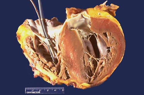
Although hypertrophy in HCM may involve any portion of the ventricles, asymmetric hypertrophy of the ventricular septum is most commonly found (approximately 90% of cases). Less often, the hypertrophy involves the ventricular walls symmetrically or is localized to the free wall of the LV.
The following shows marked left ventricular hypertrophy, with asymmetric bulging of a very large interventricular septum into the left ventricular chamber:
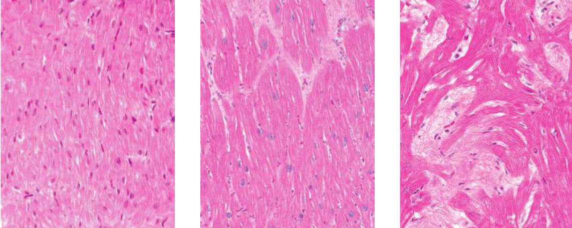
Microscopically, unlike ventricular hypertrophy resulting from hypertension or aortic stenosis in which the myocytes enlarge uniformly and remain orderly, the histology of HCM is unusual. The myocardial fibers are in a pattern of disarray. Hypertrophied fibers are oriented in chaotic directions and are surrounded by numerous fibroblasts and extracellular matrix. This myocyte disarray and fibrosis are characteristic of HCM and play a role in the abnormal diastolic stiffness and the arrhythmias common to this disorder.
The first image shows normal myocardium, the second shows hypertrophy due to hypertension, and the third shows hypertrophic cardiomyopathy with myocyte disarray and fibrosis:
Pathophysiology
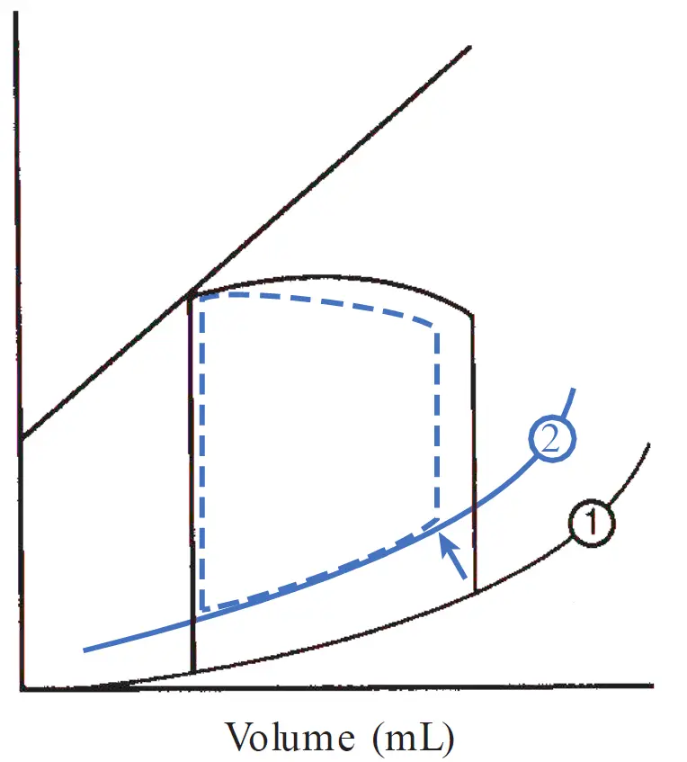
The predominant feature of HCM is marked ventricular hypertrophy that reduces the compliance and diastolic relaxation properties of the chamber, such that filling becomes impaired.
The reduced ventricular compliance alters the normal pressure–volume relationship, causing the passive diastolic filling curve to shift upward. The associated rise in diastolic LV pressure is transmitted backward, leading to elevated left atrial, pulmonary venous, and pulmonary capillary pressures. Dyspnea, especially during exertion, is thus a common symptom in this disorder.
Patients who have hypertrophy of the proximal interventricular septum may display additional findings related to transient obstruction of left ventricular outflow during systole.
HCM with Outflow Obstruction
Slightly over one third of patients with HCM have systolic outflow tract obstruction. One factor that contributes to this obstruction is hypertrophy of the proximal interventricular septum, causing physical stenosis in the left ventricular outflow tract.
Another factor contributing to the obstruction is the abnormal motion of the anterior mitral valve leaflet toward the LV outflow tract where the thickened septum protrudes. The process can be explained as follows:
-
As the left ventricle contracts, blood is forced through the narrowed outflow tract at high velocity, creating a local low-pressure zone (the Venturi effect) along with hydrodynamic drag.
-
These forces pull the tip of the anterior mitral valve leaflet toward the thickened interventricular septum during mid-to-late systole.
-
When the mitral leaflet contacts the septum, it momentarily blocks the exit path into the aorta and frequently causes secondary mitral regurgitation.
The elevated ventricular systolic pressure increases wall stress and myocardial oxygen consumption, which can result in angina. The mitral regurgitation that develops may worsen symptoms of dyspnea and contribute to left atrial stretch and the development of atrial fibrillation.
The systolic pressure gradient observed in obstructive HCM is dynamic in that its magnitude varies during contraction and depends, at any given time, on the distance between the anterior leaflet of the mitral valve and the hypertrophied septum. Situations that decrease LV cavity size (e.g. reduced venous return owing to intravascular volume depletion) bring the mitral leaflet and septum into closer proximity and promote obstruction. Conversely, conditions that increase EDV increase the distance between the anterior mitral leaflet and septum and reduce the obstruction. Positive inotropic drugs also force the mitral leaflet and septum into closer proximity and contribute to obstruction, whereas negative inotropic drugs (e.g. β-blockers, verapamil) have the opposite effect.
Although dynamic systolic outflow tract obstruction creates an impressive murmur and receives great attention, the symptoms of obstructive HCM appear to primarily stem from the increased LV stiffness and diastolic dysfunction that are also present in the nonobstructive form.
Clinical findings
The symptoms of HCM vary widely, from none to marked physical limitations. The average age of presentation is the mid-20s. The most frequent symptom is dyspnea owing to elevated diastolic LV (and therefore pulmonary capillary) pressure. This symptom is further exacerbated by the mitral regurgitation found in patients with outflow obstruction.
Angina is often a complaint of patients with HCM, even in the absence of coronary artery disease. Myocardial ischemia may be contributed to by (1) the high oxygen demand of the increased muscle mass and (2) the narrowed small branches of the coronary arteries within the hypertrophied ventricular wall. If outflow tract obstruction is present, the high systolic ventricular pressure increases myocardial oxygen demand because of the increased wall stress and contributes to ischemia.
Syncope in HCM may result from cardiac arrhythmias that arise because of the structurally abnormal myofibers. In patients with outflow tract obstruction, syncope may also be induced by exertion, as the obstruction is made worse by the increased force of contraction (i.e. due to sympathetic activation), causing a transient fall in cardiac output. Orthostatic light-headedness is also common in patients with outflow tract obstruction. This occurs because venous return to the heart is reduced on standing by the gravitational pooling of blood in the lower extremities, decreasing the left ventricular cavity size and increasing the obstruction, reducing cardiac output and cerebral perfusion.
Just like in aortic stenosis, atrial fibrillation is not well tolerated because the loss of the normal atrial “kick” impairs diastolic filling and can therefore worsen symptoms of pulmonary congestion.
Of greatest concern, the first clinical manifestation of HCM may be ventricular fibrillation, resulting in sudden cardiac death, particularly in young adults with HCM during strenuous physical exertion. Risk factors for sudden death include a history of syncope, a family history of sudden death, certain high-risk HCM mutations, and extreme hypertrophy of the LV wall ( mm in thickness).
Note
The incidence of sudden death in HCM is 2% to 4% per year in adults and 4% to 6% in children and adolescents depending on the specific mutation.
Physical Examination
A patient with mild HCM may have a normal cardiac examination. Otherwise, a common finding is an S4, generated by left atrial contraction into the stiffened LV. The forceful atrial contraction may also result in a palpable presystolic impulse over the cardiac apex (a “double apical impulse”).
Other findings are typical in patients with systolic outflow obstruction. The carotid pulse rises briskly in early systole but then quickly declines as obstruction to cardiac outflow appears.
The characteristic systolic murmur of LV outflow obstruction is rough and crescendo–decrescendo in shape, best heard at the left lower sternal border, and unlike AS, does not typically radiate to the carotid arteries. In addition, as the stethoscope is moved toward the apex, the holosystolic blowing murmur of accompanying mitral regurgitation may be auscultated.
Bedside maneuvers that alter preload and afterload can affect the intensity of the LV outflow obstruction and therefore intensify the murmur. These can help differentiate this murmur from the murmur of AS.
A commonly used technique is the Valsalva maneuver, produced by asking the patient to “bear down” (i.e. forceful exhalation with the nose, mouth, and glottis closed). This increases intrathoracic pressure, decreasing venous return and transiently reducing LV cavity size. This brings the hypertrophied septum and anterior leaflet of the mitral valve into closer proximity, creating greater obstruction, and increasing the murmur intensity. In contrast, the murmur of AS decreases in intensity during Valsalva because the decreased preload reduces the flow across the stenotic valve.
Conversely, a sudden change from standing to a squatting position increases venous return to the heart while simultaneously increasing the systemic vascular resistance. The increased preload increases the stroke volume and so increases the intensity of the AS murmur. In contrast, the increase in LV size during squatting reduces the LV outflow tract obstruction in HCM and softens the intensity of that murmur.

Treatment
β-Blockers are standard therapy for HCM because they reduce myocardial oxygen demand by slowing the heart rate and force of contraction; lessen any LV outflow gradient during exercise by reducing the force of contraction (allowing the chamber size to increase, thus separating the anterior leaflet of the mitral valve from the ventricular septum); increase passive diastolic ventricular filling time owing to the decreased heart rate; and decrease the frequency of ventricular ectopic beats.
Despite their antiarrhythmic effect, β-blockers do not prevent sudden arrhythmic death in this condition, nor have they been shown to slow disease progression.
Certain calcium channel antagonists (e.g. verapamil) may have beneficial effects on ventricular relaxation and filling and are sometimes useful in improving exercise capacity in patients who fail to respond to β-blockers. Patients who develop pulmonary congestion may benefit from mild diuretic therapy, but these drugs must be administered cautiously to avoid volume depletion, which decreases LV size and could exacerbate outflow tract obstruction. Vasodilators (including nitrates) similarly reduce LV size and should be avoided.
Atrial fibrillation in HCM should be controlled aggressively, most commonly with antiarrhythmic drugs. Effective and useful antiarrhythmic drugs for atrial fibrillation in HCM include amiodarone and disopyramide (a class IA antiarrhythmic drug that also possesses negative inotropic properties). Digitalis should be avoided in HCM because its positive inotropic effect can worsen LV outflow tract obstruction.
Sudden cardiac death has a propensity to occur in patients with HCM in association with physical exertion; therefore, strenuous exercise and competitive sports should be avoided. Although amiodarone may reduce the frequency of ventricular arrhythmias, high risk HCM patients (i.e. family history of sudden death, extreme hypertrophy with ventricular wall thickness mm, unexplained prior syncopal episodes, history of ventricular tachyarrhythmias) should receive an ICD.
Some studies have shown clinical improvement when patients with the obstructive form of HCM are treated with a dual-chamber permanent pacemaker, the two electrodes of which are placed in the right atrium and RV. The LV outflow gradient may become reduced by this procedure, possibly by altering the normal sequence of ventricular contraction, such that septal–mitral valve apposition becomes less prominent. However, this technique seems to be useful for only a small percentage of markedly symptomatic patients.
For cases with refractory symptoms, surgical therapy (myomectomy) is the “gold standard” treatment approach. This procedure involves excision of portions of the hypertrophied septal muscle mass, which usually improves outflow tract obstruction, symptoms, and exercise capacity. A less invasive alternative in select patients is percutaneous septal ablation, performed in the cardiac catheterization laboratory, in which ethanol is injected directly into the first major septal coronary artery (a branch of the left anterior descending artery), causing a small, controlled myocardial infarction. The desired and often achieved result is reduction of septal thickness and improvement in outflow tract obstruction.
Theoretically, infective endocarditis could develop in patients with the obstructive form of HCM. However, that is rare and routine antibiotic prophylaxis for prevention of endocarditis prior to high risk procedures is not recommended in this condition by current US guidelines.
Finally, genetic counseling should be provided to all patients with HCM. Because it is an autosomal dominant disease, children of affected persons have a 50% chance of inheriting the abnormal gene. First-degree relatives of patients with HCM should be screened by physical examination, ECG, echocardiography, and sometimes genetic testing. Even asymptomatic HCM patients are at risk of complications, including sudden death.
Restrictive cardiomyopathy
Restrictive cardiomyopathies are less common than DCM and HCM. They are characterized by abnormally stiff (but not necessarily thickened) ventricles with impaired diastolic filling but usually normal, or near normal, systolic function. This condition results from either fibrosis or scarring of the endocardium or infiltration of the myocardium by an abnormal substance.
The most common cause of restrictive cardiomyopathy in non-tropical countries is amyloidosis, which is a systemic disease in which insoluble misfolded proteins deposit within tissues, causing organ dysfunction. Amyloid deposition is diagnosed histologically by the Congo red stain, which reveals amyloid fibrils with a characteristic green birefringence under polarized light.
Amyloid fibrils can develop from several different proteins that distinguish the categories of disease. Primary amyloidosis is caused by deposition of immunoglobulin light chain AL fragments secreted by a plasma cell tumors (i.e. monoclonal gammopathy). In contrast, secondary amyloidosis is characterized by the presence of AA amyloid, prominent in a variety of chronic inflammatory conditions, such as rheumatoid arthritis.
Less common is hereditary amyloidosis, an autosomal dominant condition in which amyloid fibrils form from mutations in the transthyretin protein. Senile amyloidosis refers to a condition in the elderly, in which amyloid deposits, derived from transthyretin or other proteins, are found scattered throughout the vascular system, muscles, kidney, and lung.
In each form of amyloidosis, cardiac involvement is marked by deposition of extracellular amyloid between myocardial fibers in the atria and ventricles, in the coronary arteries and veins, and in the heart valves.
Note
AA (secondary) amyloidosis rarely causes clinically significant cardiac involvement compared to AL (light chain) and ATTR (transthyretin) amyloidosis.
Reduced compliance of the ventricles results in an upward shift of the passive ventricular filling curve, leading to abnormally high diastolic pressures. This results in an elevated systemic and pulmonary venous pressures, causing signs of right- and left-sided vascular congestion. This also causes decreased ECV and preload, decreasing stroke volume and forward cardiac output.
Other causes of restrictive cardiomyopathy include hemochromatosis, glycogen storage diseases, sarcoidosis, scleroderma, endomyocardial fibrosis, hypereosinophilic syndrome, metastatic tumors, and radiation therapy.
Clinical manifestations
It follows from the underlying pathophysiology that signs of left- and right-sided heart failure are expected. Decreased cardiac output is manifested by fatigue and decreased exercise tolerance.
Systemic congestion (often more prominent than pulmonary congestion in this syndrome) leads to jugular venous distention, peripheral edema, and ascites with a large, tender liver.
Orthostatic hypotension is present in about 10% of patients, likely contributed to by amyloid deposition in the autonomic nervous system and peripheral blood vessels. Infiltration of amyloid into the cardiac conduction system can cause arrhythmias and conduction impairments, which can result in syncope or sudden death.
Physical signs and diagnostic tests
Signs of congestive heart failure are often present, including pulmonary rales, distended neck veins, ascites, and peripheral edema. Similar to constrictive pericarditis, jugular venous distention may paradoxically worsen with inspiration (the Kussmaul sign) because the stiffened RV cannot accommodate the increased venous return.
Chest radiography usually shows a normal-sized heart with signs of pulmonary congestion.
ECG often displays non-specific ST and T wave abnormalities; conduction disturbances such as atrioventricular block or a bundle branch block may be present.
Restrictive cardiomyopathies share nearly identical symptoms, physical signs, and hemodynamic profiles with constrictive pericarditis. However, it is important to distinguish these two entities because constrictive pericarditis is often correctable, whereas interventions for restrictive cardiomyopathies are more limited. The most useful diagnostic tools to differentiate the two conditions are transvenous endomyocardial biopsy, CT, and MRI.
For example, in restrictive cardiomyopathies, transvenous endomyocardial biopsy may demonstrate the cause of the condition (e.g. presence of amyloid fibrils in amyloidosis, or iron deposits in hemochromatosis). CT or MRI scans accurately identify the thickened pericardium present in most patients with constrictive pericarditis, a finding that is not present in restrictive cardiomyopathy.
Treatment
Restrictive cardiomyopathy typically has a very poor prognosis, except when treatment can target the underlying cause. For example, phlebotomy and iron chelation therapy may be helpful in hemochromatosis.
Symptomatic therapy for all etiologies includes salt restriction and cautious use of diuretics to improve symptoms of congestion. Unlike the dilated cardiomyopathies, vasodilators are not helpful because systolic function is usually preserved. Maintenance of sinus rhythm (e.g. converting atrial fibrillation if it occurs) is important to maximize diastolic filling and forward cardiac output.
Some restrictive cardiomyopathies are prone to intraventricular thrombus formation, warranting chronic oral anticoagulant therapy. In the case of primary (AL) amyloidosis, chemotherapy followed by autologous bone marrow stem cell transplantation has proved effective in selected patients with early cardiac involvement.
Other forms of cardiomyopathy
Noncompaction cardiomyopathy
Left ventricular noncompaction (LVNC), also referred to as spongiform Cardiomyopathy, is a rare condition with features that overlap with hypertrophic, restrictive and dilated cardiomyopathies. Normally during embryogenesis, the developing myocardial fibers form an interwoven meshwork which has spongy consistency. This mesh later undergoes compaction and transforms to the solid musculature found in normal heart. When this process is defective, it results in Non-compaction cardiomyopathy.
The exact cause of this arrest in development is not understood, but up to 50% of patients with this condition have affected family members, and autosomal dominant, autosomal recessive and X-linked patterns of inheritance have been found. Mutations in at least nine genes encoding sarcomere proteins have been identified in patients with LVNC (including mutations that have also been associated with hypertrophic and dilated cardiomyopathies), resulting in either the isolated disorder or a syndrome with other forms of congenital heart disease.
Ventricular trabeculation is excessively prominent as can be seen in the following image.
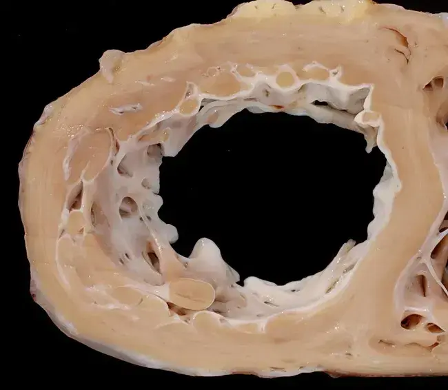
The abnormal regions of myocardium typically contract poorly. Patients with this condition may present in childhood or adulthood with symptoms of heart failure (due to systolic and/ or diastolic dysfunction), ventricular arrhythmias, or thromboembolism.
The diagnosis is usually established by typical features on 2-dimensional and Doppler echocardiography or by cardiac MRI. The prognosis is variable, but is worse among symptomatic patients compared to those found to have the disorder incidentally by imaging studies.
Management is aimed at treating the symptoms and complications of LVNC as there is no corrective therapy for the underlying condition itself. Depending on the clinical manifestations, approaches may include standard treatment of heart failure, ICD implantation or management of life-threatening ventricular arrhythmias, chronic anticoagulation for those with accompanying atrial fibrillation or significant contractile dysfunction to prevent thromboembolism, and cardiac transplantation for those with advanced, refractory heart failure.
Arrhythmogenic Right Ventricular Cardiomyopathy
ARVC, also termed arrhythmogenic right ventricular dysplasia, is another genetic form of cardiomyopathy. It is characterized by replacement of right ventricular (and occasionally left ventricular) myocardium with adipose and fibrous tissue, resulting in rhythm disturbances and contractile dysfunction.
Ventricular arrhythmias originating from the abnormal ventricle are common and may result in palpitations, syncope, and even sudden cardiac death. Symptoms often begin in the teen years, and ARVC is another cause of sudden death among young athletes.
Both autosomal dominant and recessive inheritance forms of ARVC have been identified. The majority of mutations occur in genes that encode components of desmosomes, leading to fibrosis and aberrant signaling with proliferation of adipose tissue in the myocardium.
Transvenous endomyocardial biopsy of the RV may demonstrate fatty or fibro fatty replacement of the myocardium, but has a high false negative rate for identifying the disorder. Thus, diagnostic criteria also rely on family history, ECG abnormalities, the presence of arrhythmias emanating from the RV, morphologic abnormalities detected by imaging studies (especially cardiac MRI), and genetic testing for specific mutations.
Management typically includes ICD implantation because the disease is progressive and life-threatening ventricular tachycardia is common.
See also
References
Additional Reading
- Ramnani DM. Non-Compaction Cardiomyopathy. WebPathology. Accessed July 29, 2026. https://www.webpathology.com/images/cardiovascular/heart/cardiomyopathies/58005
- Arbustini E, Narula J, Tavazzi J, et al. The MOGE(S) classification of cardiomyopathy for clinicians. J Am Coll Cardiol. 2014;64:304–318.
- Bhatia NL, Tajik AJ, Wilansky S, et al. Isolated noncompaction of the left ventricular myocardium in adults: A systematic overview. J Card Fail. 2011;17:771–778.
- Elkayam U. Clinical characteristics of peripartum cardiomyopathy in the United States: Diagnosis, prognosis, and management. J Am Coll Cardiol. 2011;58:659–670.
- Gersh BJ, Maron BJ, Bonow RO, et al. 2011 ACCF/ AHA guideline for the diagnosis and treatment of hypertrophic cardiomyopathy: Executive summary. J Am Coll Cardiol. 2011;58:2703–2738.
- Guan J, Mishra S, Falk RH, et al. Current perspectives on cardiac amyloidosis. Am J Physiol Heart Circ Physiol. 2012;302:H544–H552
- Maron BJ, Ommen SR, Semsarian C, et al. Hypertrophic cardiomyopathy: Present and future, with translation into contemporary cardiovascular medicine. J Am Coll Cardiol. 2014;64:83–99
- Maron BJ, Towbin JA, Thiene G, et al. Contemporary definitions and classification of the cardiomyopathies: An American Heart Association scientific statement from the Council on Clinical Cardiology, Heart Failure and Transplantation Committee; Quality of Care and Outcomes Research and Functional Genomics and Translational Biology Interdisciplinary Working Groups; and Council on Epidemiology and Prevention. Circulation. 2006;113:1807–1816.
- Murray B. Arrhythmogenic right ventricular dysplasia/ cardiomyopathy (ARVD/ C): A review of molecular and clinical literature. J Genet Couns. 2012;21:494–504
- Sturm AC. Genetic testing in the contemporary diagnosis of cardiomyopathy. Curr Heart Fail Rep. 2013;10:63–72