Part 8: Heart Failure
Mon Jul 27 2026
By B. Hassan
The heart normally accepts blood at low filling pressures during diastole and then propels it forward at higher pressures in systole. Heart failure is present when the heart is unable to pump blood forward at a sufficient rate to meet the body’s metabolic demands or is able to do so only if cardiac filling pressures are abnormally high. Although extra-cardiac causes may meet this definition through inadequate tissue perfusion (e.g. severe hemorrhage) or through increased metabolic demands (e.g. hyperthyroidism), in this part, we only consider cardiac causes.
Heart failure may be the final and most severe manifestation of nearly every form of cardiac disease, including coronary atherosclerosis, myocardial infarction, valvular diseases, hypertension, congenital heart disease, and the cardiomyopathies. The number of patients with heart failure is increasing, not only because the population is aging but also because of interventions that prolong survival after heart diseases such as myocardial infarction. As a result, heart failure now is the most common diagnosis of hospitalized patients aged 65 years and older.
Physiology
Experimental studies of isolated muscle segments have revealed an important principle that can be generalized to the intact heart. As a muscle segment is stretched, the relation between its length and the tension it passively develops is curvilinear, reflecting its intrinsic elastic properties. If the muscle is first passively stretched and then stimulated while its ends are held at fixed positions (termed isometric contraction), the total tension generated is proportional to the length of the muscle at the time of stimulation. This is because stretching the muscle before stimulation optimizes the overlap and interaction between myosin and actin filaments, increasing the number of cross bridges and the force of contraction.
This relationship between the initial fiber length and force development is of great importance in the intact heart: within a physiologic limit, the larger the end-diastolic volume (EDV), the more the fibers are stretched before stimulation and the greater the force of contraction it generates. This is the basis of the Frank–Starling relationship, the observation that ventricular output increases in relation to the preload.
A second observation arises when the fibers are not held at a fixed length but are allowed to shorten against a fixed load (termed the afterload). In this situation (termed an isotonic contraction), once the tension inside the muscle matches the load, the muscle begins to shorten (i.e. the weight moves when the force generated by the muscle reaches the force of the weight). It was found that if the fibers are stretched to a longer length before stimulation but the afterload is kept constant, the muscle will shorten a greater distance, but the final length of the muscle after shortening only depends on the afterload, not the initial length of the muscle.
Suppose a muscle with an initial length of 8cm lifts a 10 N weight, and after stimulation, the muscle’s final length shortens to 6cm. Then if we stretch the muscle such that its initial length is 10cm while keeping the same afterload, then after stimulation, the final length would still be 6cm. Notice that increasing the stretch makes the muscle shorten a greater distance (i.e. higher force of contraction) due to the Frank–Starling relationship, but the final length after shortening only depends on the afterload and not the initial length; the higher the afterload, the higher the length of the muscle after contraction. In other words, the greater the load opposing contraction, the less the muscle fiber can shorten.
This concept of afterload can also be generalized to the intact heart. The size of the chamber at the end of each contraction (i.e. end-systolic volume) depends on the load against which the ventricle contracts but is independent of the stretch on the myocardial fibers before contraction.
A third key experimental observation relates to myocardial contractility, which can be influenced by chemical and hormonal influences such as exposure to catecholamines. When contractility is enhanced pharmacologically, the relation between initial fiber length and force development is shifted upward such that a greater total tension develops with isometric contraction at any given preload. Similarly, when contractility is increased and the cardiac muscle is allowed to shorten against a fixed afterload, the fiber contracts to a greater extent and achieves a shorter final length compared with the baseline state.
Pressure–Volume Loop
A useful graphic display to illustrate the determinants of cardiac function is the ventricular pressure–volume loop, which relates changes in ventricular volume to corresponding changes in pressure throughout the cardiac cycle. We described the pressure volume loop previously when talking about the cardiac cycle.
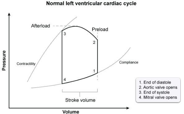
As the volume increases during diastole, it is associated with a small rise in pressure, in accordance with compliance of the myocardium, reflected by the lower curve in the graph.
Next, left ventricular systolic contraction causes the ventricular pressure to rise. When the pressure in the LV exceeds that of the left atrium (point 1), the mitral valve is forced to close. The volume at point 1 is the end diastolic volume.
As the pressure continues to increase, the ventricular volume does not immediately change, because the aortic valve has not yet opened; therefore, this phase is called isovolumetric contraction. The pressure against which the ventricle ejects (a component of afterload) is represented by the curve between 2 and 3.
As the ventricle continues to relax, its pressure declines while its volume remains constant because the mitral valve has not yet opened. This phase is known as isovolumetric relaxation.
At point 3, the contraction stops, and the aortic valve slams shut, producing S2. The volume at this point is the End Systolic Volume (ESV), which represents the blood left in the heart after full ventricular contraction.
Contractile function of the heart
In a healthy person, cardiac output (CO) is matched to the body’s total metabolic need. CO is proportional to both the stroke volume (SV, meaning the volume of blood ejected in each contraction) and the heart rate (HR):
As we discussed above, the 3 major determinants of stroke volume are preload, afterload, and myocardial contractility.
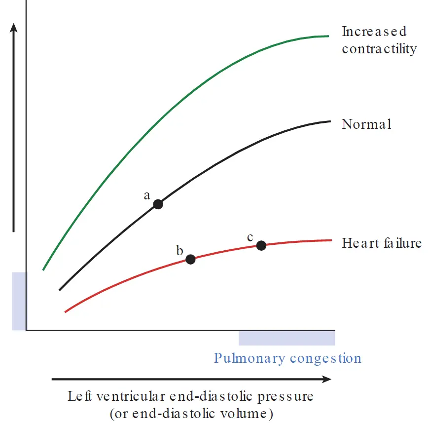
The preload relates to the EDV. The more the ventricle is distended during diastole, the greater the volume ejected during the next systole. This is just the Frank-Starling relationship, and can be illustrated graphically by the Frank–Starling curve, also known as the ventricular function curve. The graph relates a measurement of cardiac performance (e.g. CO or SV) on the y-axis as a function of preload on the x-axis.
Conditions that decrease intravascular volume, and thereby reduce ventricular preload (e.g. dehydration or severe hemorrhage), result in a smaller EDV and hence a reduced stroke volume. Conversely, an increased EDV (e.g. a large IV fluid infusion) results in a greater-than-normal stroke volume.
The following is a Frank–Starling curve. For a normal heart, cardiac performance continuously increases as a function of preload. States of increased contractility (e.g. norepinephrine infusion) are characterized by an increased cardiac performance at any level of preload. Conversely, decreased contractility (i.e. heart failure) is characterized by a curve that is shifted downwards. Point a is an example of a normal person at rest. Point b represents the same person after developing heart failure: stroke volume has fallen, and the decreased LV emptying results in elevation of the EDV. Further increase in LV filling (e.g. increased circulating volume) in the heart failure patient is represented by point c, which resides on the relatively flat part of the curve: stroke volume is only slightly increased with any increase in preload, but the significantly increased EDV results in blood stagnation and pulmonary congestion.
Afterload in the intact heart reflects the resistance that the ventricle must overcome to eject its contents. It is more formally defined as the ventricular wall stress that develops during systolic ejection. Wall stress (), like pressure, is expressed as force per unit area and, for the left ventricle, it may be estimated from Laplace relationship:
where:
- is ventricular pressure
- is ventricular chamber radius
- is ventricular wall thickness
Thus, ventricular wall stress increases in response to a higher pressure load (e.g. hypertension) or an increased chamber size (e.g. a dilated left ventricle). Conversely, an increase in wall thickness () serves a compensatory role in reducing wall stress, because the force is distributed over a greater mass per unit surface area of ventricular muscle.
Alterations in Preload
If afterload and contractility are held constant but preload increases (e.g. by administration of IV fluids), left ventricular EDV rises. This increases the stroke volume via the Frank–Starling mechanism such that the end-systolic volume (ESV) achieved is the same as it was before increasing the preload. This means that the normal left ventricle is able to adjust its stroke volume and effectively empty its contents to match its diastolic filling volume, as long as contractility and afterload are kept constant.
Although EDV and end-diastolic pressure (EDP) are often used interchangeably as markers of preload, the relationship between filling volume and pressure (i.e. ventricular compliance) largely governs the extent of ventricular filling. If ventricular compliance is reduced (e.g. severe LV hypertrophy), it reduces the ability of the chamber to fill during diastole, resulting in a lower-than-normal EDV, while ESV remains unchanged, thus stroke volume (EDV - ESV) is reduced
Alterations in Afterload
If preload and contractility are held constant and afterload increases (e.g. hypertension or aortic stenosis), the pressure generated by the left ventricle during ejection increases. In this situation, more ventricular work is expended in overcoming the resistance to ejection and therefore less fiber shortening takes place, resulting in a greater-than-normal ESV, thus stroke volume (EDV-ESV) is reduced. The dependence of the ESV on afterload is approximately linear: the greater the afterload, the higher the ESV.
Putting it all graphically
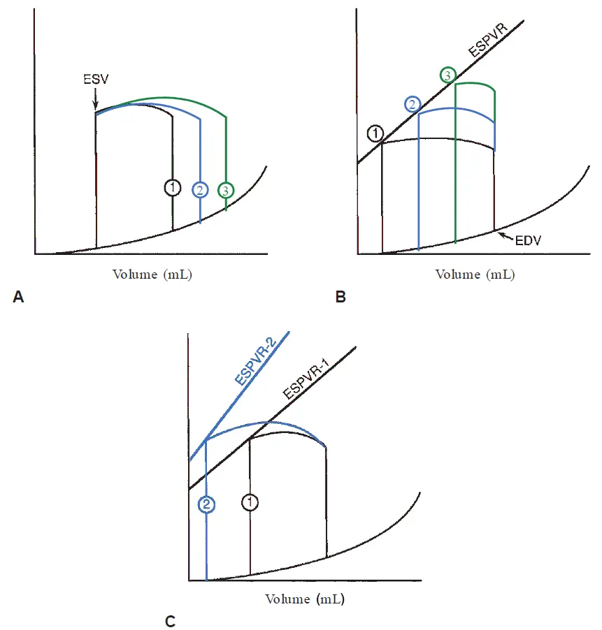
For graph A, when arterial pressure (afterload) and contractility are held constant, sequential increases in preload measured as EDV (lines 1, 2, and 3) are associated with loops that have progressively higher stroke volumes but a constant ESV.
For graph B, when the preload (EDV) and contractility are held constant, sequential increases (points 1, 2, and 3) in afterload are associated with loops that have progressively lower stroke volumes and higher end-systolic volumes. There is a nearly linear relationship between the afterload and ESV, termed the end-systolic pressure–volume relation (ESPVR).
For graph C, a positive inotropic intervention shifts the ESPVR upward and leftward from ESPVR-1 to ESPVR-2, resulting in loop 2, which has a larger stroke volume and a smaller ESV than the original loop 1.
Pathophysiology
Chronic heart failure may result from a wide variety of etiologies, these can be grouped into those that impair ventricular contractility, increase afterload, or impair ventricular relaxation and filling.
Heart failure that results from an abnormality in ventricular emptying (due to impaired contractility or greatly excessive afterload) is termed systolic dysfunction, whereas heart failure caused by abnormalities of diastolic relaxation or ventricular filling is termed diastolic dysfunction. However, there is much overlap, and many patients demonstrate both systolic and diastolic abnormalities.
It is common to categorize heart failure into 2 general categories based on the ejection fraction (EF). These are heart failure with reduced EF (i.e. primarily systolic dysfunction) and heart failure with preserved EF (i.e. primarily diastolic dysfunction). In the United States, approximately one half of patients with heart failure fall into each of these categories.
Heart Failure with Reduced EF (HFrEF)
In states of systolic dysfunction, the affected ventricle has a diminished capacity to eject blood because of impaired myocardial contractility or pressure overload (i.e. excessive afterload).
The following depicts the effects of systolic dysfunction due to impaired contractility on the pressure–volume loop. The normal pressure–volume loop (solid line) is compared with one demonstrating systolic dysfunction (dashed blue line). In HFrEF, the ESPVR is shifted downward and rightward (from line 1 to line 2) such that systolic emptying ceases at a higher-than-normal ESV (ESV increases, depicted by the arrow).
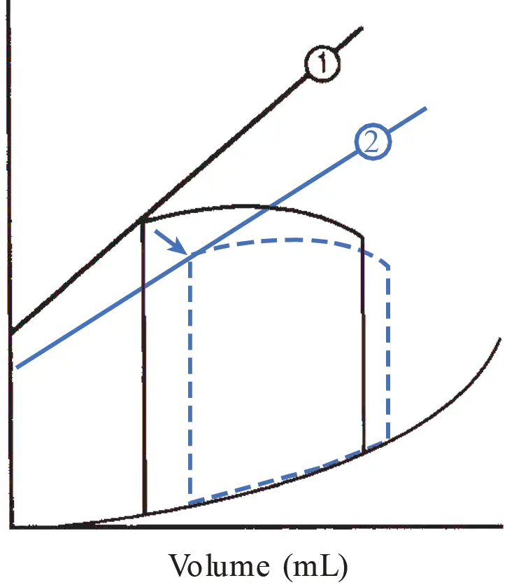
When normal pulmonary venous return is added to the increased ESV from the previous cycle (i.e. due to incomplete emptying), the higher-than-normal EDV (preload) induces a compensatory rise in stroke volume (via the Frank–Starling mechanism), but impaired contractility and the reduced EF cause the ESV to remain elevated.
During diastole, the persistently elevated LV pressure is transmitted to the left atrium and to the pulmonary veins and capillaries. An elevated pulmonary capillary hydrostatic pressure (usually greater than 20 mmHg) results in the transudation of fluid into the pulmonary interstitium causing symptoms of pulmonary edema.
Heart Failure with Preserved EF (HFpEF)
Patients who exhibit heart failure with preserved EF frequently demonstrate abnormalities of ventricular diastolic function: impaired early diastolic relaxation (an active, energy-dependent process), increased stiffness of the ventricular wall (a passive property), or both.
Acute myocardial ischemia is an example of a condition that transiently inhibits energy delivery and diastolic relaxation. Conversely, left ventricular hypertrophy, fibrosis, or restrictive cardiomyopathy causes the LV walls to become chronically stiffened. Certain pericardial diseases (e.g. cardiac tamponade) present an external force that limits ventricular filling and represent potentially reversible forms of diastolic dysfunction.
The following shows the effect of impaired diastolic function on the pressure–volume loop (dashed blue line). The compliance curve is shifted upward (from line 1 to line 2) such that at any diastolic volume, the ventricular pressure is higher than normal. The result is a decreased EDV (arrow) because of reduced filling of the stiffened ventricle at a higher-than-normal end-diastolic pressure.
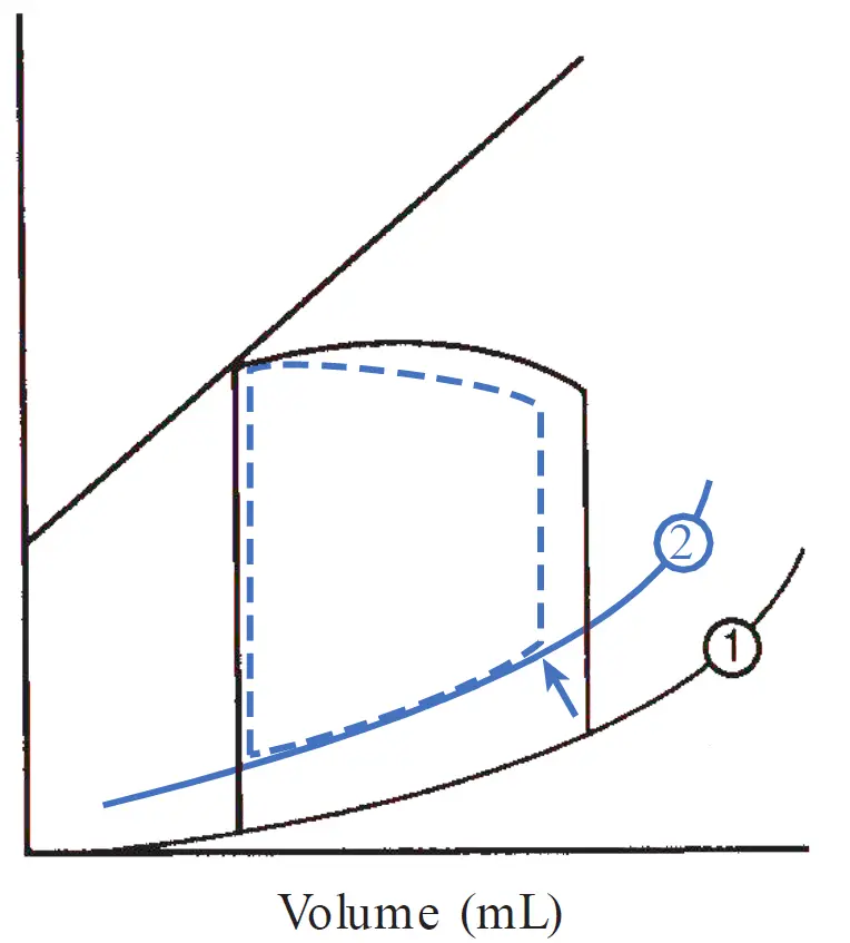
Patients with diastolic dysfunction often manifest signs of vascular congestion because the elevated diastolic pressure is transmitted retrograde to the pulmonary circulation.
Right-Sided Heart Failure
Whereas the physiologic principles described above may be applied to both right-sided and left-sided heart failure, there are distinct differences between the two ventricles. Compared with the left ventricle, the right ventricle (RV) is a thin-walled, highly compliant chamber that accepts its blood volume at low pressures and ejects against a low pulmonary vascular resistance.
As a result of its high compliance, the RV has little difficulty accepting a wide range of filling volumes without marked changes in filling pressure. Conversely, the RV is quite susceptible to failure in situations that present a sudden increase in afterload, such as pulmonary embolism.
The most common cause of right-sided heart failure is actually the presence of left-sided heart failure. In this situation, the LV congestion is transmitted to the LA and to the pulmonary circulation, increasing the afterload of the RV. Isolated right heart failure is less common and usually reflects increased RV afterload owing to primary lung diseases. Right-sided heart disease that results from a primary pulmonary pathology is known as cor pulmonale.
When the RV fails, the elevated diastolic pressure is transmitted retrograde to the right atrium with subsequent congestion of the systemic veins. Indirectly, isolated right heart failure may also influence left heart function: the decreased right ventricular output reduces blood return to the LV (i.e., diminished preload), causing left ventricular stroke volume to decline.
Cardiac causes of right-sided heart failure:
- Left-sided heart failure
- Pulmonic valve stenosis
- Right ventricular infarction
Pulmonary parenchymal diseases causing right-sided heart failure:
- Chronic obstructive pulmonary disease
- Interstitial lung disease (e.g., sarcoidosis)
- Chronic lung infection or bronchiectasis
Pulmonary vascular diseases causing right-sided heart failure:
- Pulmonary embolism
- Pulmonary hypertension
Compensatory mechanisms in heart failure
Frank–Starling Mechanism
Heart failure caused by impaired left ventricular contractile function causes a downward shift of the ventricular contractility curve. Consequently, at a given preload, stroke volume decreases compared to normal. This results in incomplete chamber emptying, so that the volume of blood that accumulates in the ventricle during diastole (EDV) is higher than normal.
This increased stretch on the myofibers, acting via the Frank–Starling mechanism, induces a greater stroke volume on subsequent contraction, which helps to empty the enlarged LV and preserve cardiac output. This beneficial compensatory mechanism has its limits, however. In the case of severe heart failure, the curve may be nearly flat at higher diastolic volumes, reducing the increase in cardiac output achieved by the increased chamber filling.
Neurohormonal Compensation
Several important neurohormonal compensatory mechanisms are activated in heart failure in response to the decreased cardiac output. Some of the most important ones are the adrenergic nervous system and the renin–angiotensin–aldosterone system.
In part, these mechanisms serve to increase systemic vascular resistance, which helps maintain arterial perfusion to vital organs, even in the setting of a reduced cardiac output. That is, because blood pressure (BP) is equal to the product of cardiac output (CO) and total peripheral resistance (TPR):
A rise in TPR induced by these compensatory mechanisms can nearly balance the fall in CO and, in the early stages of heart failure, maintain fairly normal BP. In addition, neurohormonal activation results in salt and water retention, which increases intravascular volume and left ventricular preload, maximizing stroke volume via the Frank–Starling mechanism. Although the acute effects of neurohormonal stimulation may be beneficial, chronic activation of these mechanisms often ultimately proves detrimental to the failing heart and contributes to a progressive downhill course.
Adrenergic Nervous System
The fall in cardiac output in heart failure is sensed by baroreceptors in the carotid sinus and aortic arch. These receptors decrease their rate of firing in proportion to the fall in BP, and the signal is transmitted by the 9th (glossopharyngeal) and 10th (vagus) cranial nerves to the cardiovascular center in the medulla. As a consequence, sympathetic outflow to the heart and peripheral circulation is enhanced, and parasympathetic tone is diminished.
The immediate consequences are: increased heart rate, increased ventricular contractility, and vasoconstriction of systemic vessels via α-receptor stimulation. The increased heart rate and ventricular contractility directly increase cardiac output. Vasoconstriction of the peripheral circulations is also initially beneficial. Venous constriction augments blood return to the heart, which increases preload and raises stroke volume through the Frank–Starling mechanism, as long as the ventricle is operating on the ascending portion of its ventricular performance curve.
Arteriolar constriction increases the peripheral vascular resistance and thus helps maintain blood pressure (). The regional distribution of α-receptors is such that during sympathetic stimulation, blood flow is redistributed to vital organs (e.g. heart and brain) at the expense of the skin, splanchnic viscera, and kidneys.
Renin–Angiotensin–Aldosterone System (RAAS)
This system is activated early in patients with heart failure, mediated by increased renin release. The main stimuli for renin secretion from the juxtaglomerular cells of the kidney in heart failure patients include:
-
Decreased renal artery perfusion pressure secondary to low cardiac output
-
Decreased salt delivery to the macula densa of the kidney owing to alterations in intrarenal hemodynamics
-
Direct stimulation of juxtaglomerular β-receptors by the adrenergic nervous system.
Renin is an enzyme that cleaves circulating angiotensinogen to form angiotensin I, which is then rapidly cleaved by endothelial angiotensin-converting enzyme (ACE) to form angiotensin II, a potent vasoconstrictor. Angiotensin II constricts arterioles and raises total peripheral resistance, serving to maintain systemic blood pressure. In addition, angiotensin II acts to increase intravascular volume by two mechanisms:
-
At the hypothalamus, it stimulates thirst and therefore water intake
-
At the adrenal cortex, it acts to increase aldosterone secretion, which promotes sodium reabsorption from the renal distal convoluted tubules, causing salt-water retention and increasing intravascular volume.
The rise in intravascular volume increases left ventricular preload and thereby increases cardiac output via the Frank–Starling mechanism in patients on the ascending portion of the ventricular performance curve.
Failure of the compensatory mechanisms
Although each of these neurohormonal alterations is initially beneficial, continued activation ultimately proves harmful. For example, the increased intravascular volume and increased venous return may eventually worsen pulmonary vasculature congestion, exacerbating pulmonary edema symptoms. Furthermore, the elevated arteriolar resistance increases the afterload against which the failing LV contracts and may ultimately impair stroke volume and reduce cardiac output. In addition, the increased heart rate increases metabolic demand and can therefore further reduce the performance of the failing heart.
Continuous sympathetic activation results in down-regulation of cardiac β-adrenergic receptors, contributing to a decrease in the myocardium’s sensitivity to circulating catecholamines and a reduced inotropic response.
Chronically elevated levels of angiotensin II and aldosterone also provoke the production of cytokines and stimulate fibroblasts, resulting in fibrosis and adverse remodeling of the failing ventricles heart.
Because the undesired consequences of chronic neurohormonal activation eventually outweigh the benefits, much of today’s pharmacologic therapy of heart failure is designed to moderate these “compensatory” mechanisms.
Natriuretic Peptides
In contrast to the ultimately adverse consequences of the neurohormonal alterations described above, the natriuretic peptides are natural “beneficial” hormones secreted in response to the increased intra-atrial pressures in heart failure. The best studied of these are atrial natriuretic peptide (ANP) and B-type natriuretic peptide (BNP). ANP is stored in atrial cells and is released in response to atrial distention. BNP is not detected normally but is produced when ventricular myocardium is subjected to hemodynamic stress (e.g. in heart failure or myocardial infarction).
Actions of these peptides are mediated by specific natriuretic receptors and are largely opposite to those of the other hormone systems activated in heart failure. They result in excretion of sodium and water, vasodilatation, inhibition of renin secretion, and antagonism of the effects of angiotensin II on aldosterone. Although these effects are beneficial in heart failure, they are usually not sufficient to fully counteract the vasoconstriction and volume-retaining effects of the other activated hormonal systems.
Ventricular Hypertrophy and Remodeling
Ventricular hypertrophy and remodeling are important compensatory processes that develop over time in response to hemodynamic burdens. Wall stress is often increased in heart failure because of either LV dilatation (increased chamber radius) or the need to generate high systolic pressures to overcome excessive afterload (e.g. aortic stenosis or hypertension). A sustained increase in wall stress (along with neurohormonal and cytokine alterations) stimulates the development of myocardial hypertrophy.
This increased muscle mass serves as a compensatory mechanism that helps maintain contractile force and counteracts the elevated ventricular wall stress. However, because of the increased stiffness of the hypertrophied wall, these benefits come at the expense of higher-than-normal diastolic ventricular pressures (i.e. diastolic failure), which are transmitted to the left atrium and pulmonary vasculature.
Chronic chamber dilatation owing to volume overload (e.g. chronic mitral or aortic regurgitation) results in the synthesis of new sarcomeres in series with the old, causing the myocytes to elongate. The radius of the ventricular chamber enlarges in proportion to the increase in wall thickness, and is termed eccentric hypertrophy.
Chronic pressure overload (e.g. hypertension or aortic stenosis) results in the synthesis of new sarcomeres in parallel with the old (i.e. the myocytes thicken), termed concentric hypertrophy. In this situation, the wall thickness increases without proportional chamber dilatation, and wall stress may be reduced substantially.
Such hypertrophy and remodeling help reduce wall stress and maintain contractile force, but ultimately may cause further decline in ventricular function, allowing the chamber to dilate out of proportion to wall thickness. When this occurs, the excessive hemodynamic burden on the contractile units produces a downward spiral of deterioration with progressive heart failure.
Precipitating factors
Many patients with heart failure remain asymptomatic for extended periods either because the impairment is mild or because cardiac dysfunction is balanced by the compensatory mechanisms described earlier. Often, clinical manifestations are precipitated by factors that increase the cardiac workload and initiate a state of decompensation.
For example, conditions of increased metabolic demand such as fever may not be matched by a sufficient increase in output by the failing heart, so that symptoms of cardiac insufficiency are precipitated.
Tachyarrhythmias precipitate heart failure by decreasing diastolic ventricular filling time and increasing myocardial oxygen demand. Excessively low heart rates directly cause a drop in cardiac output (remember, ).
An increase in salt ingestion, renal dysfunction, or failure to take prescribed diuretic medications may cause volume overload, promoting systemic and pulmonary congestion.
Uncontrolled hypertension depresses systolic function because of excessive afterload.
A pulmonary embolism results in both hypoxemia (therefore decreased myocardial oxygen supply) and increased right ventricular afterload.
Ischemic heart disease, ethanol ingestion, or negative inotropic medications (e.g. large doses of β-blockers) can all depress myocardial contractility and precipitate symptoms in the otherwise compensated congestive heart failure patient.
Clinical Presentation
The most prominent manifestation of chronic left ventricular failure is dyspnea on exertion. Controversy regarding the cause of this symptom has centered on whether it results primarily from pulmonary venous congestion or from decreased forward cardiac output.
A pulmonary venous pressure over 20 mmHg leads to transudation of fluid into the pulmonary interstitium and congestion of the lungs. The resulting reduced pulmonary compliance increases the work of breathing. Moreover, the excess fluid in the interstitium compresses the walls of the bronchioles and alveoli, increasing the resistance to airflow and requiring greater effort of respiration. In addition, J receptors are stimulated, mediating a rapid shallow breathing pattern.
Heart failure patients can also suffer from dyspnea even in the absence of pulmonary congestion, because reduced blood flow to the overworked respiratory muscles and accumulation of lactic acid may also contribute to that sensation. Heart failure may initially cause dyspnea only on exertion, but more severe dysfunction results in symptoms at rest as well.
Other manifestations may include altered mental status due to reduced cerebral perfusion and impaired urine output during the day because of decreased renal perfusion. There is usually increased urinary frequency at night (nocturia) because, while supine, blood flow is redistributed to the kidney, promoting renal perfusion and diuresis. Reduced skeletal muscle perfusion may result in fatigue and weakness.
Other congestive manifestations include orthopnea, paroxysmal nocturnal dyspnea (PND), and nocturnal cough. Orthopnea is the development of dyspnea while lying flat that is relieved by sitting upright. It results from the redistribution of intravascular blood from the gravity-dependent portions of the body (abdomen and lower extremities) toward the circulation and to the lungs after lying down.
Note
The degree of orthopnea is generally assessed by the number of pillows on which the patient sleeps to avoid breathlessness. Sometimes, orthopnea is so significant that the patient may try to sleep upright in a chair.
PND is severe breathlessness that awakens the patient from sleep 2 to 3 hours after going to bed. This results from the gradual reabsorption of the lower extremity edema into the circulation after lying down, with subsequent volume overload and pulmonary congestion.
A nocturnal cough is another symptom of pulmonary congestion and is produced by a mechanism similar to orthopnea. Hemoptysis may result from rupture of engorged bronchial veins.
In right-sided heart failure, the elevated systemic venous pressure can result in abdominal discomfort because the liver becomes engorged and its capsule stretched. Similarly, anorexia and nausea may result from edema within the gastrointestinal tract. Peripheral edema, especially in the lower limbs, also reflect increased hydrostatic venous pressures. Because of the effect of gravity, it tends to worsen while the patient is upright and is often improved by morning after lying down at night. Even before peripheral edema develops, the patient may note an unexpected weight gain resulting from the accumulation of interstitial fluid.
The symptoms of heart failure are commonly graded according to the New York Heart Association (NYHA) classification, and patients may shift from one class to another, in either direction, over time. A newer system classifies patients according to their stage in the course of heart failure, progressing in only one direction (from A to D).
NYHA classification system:
-
I: No limitation of physical activity
-
II: Slight limitation of activity. Dyspnea and fatigue with moderate exertion (e.g. walking up stairs quickly)
-
III: Marked limitation of activity. Dyspnea with minimal exertion (e.g. slowly walking up stairs)
-
IV: Severe limitation of activity. Symptoms are present even at rest.
Stages of Chronic Heart Failure (the newer classification system):
-
A: The patient is at risk of developing heart failure but has not yet developed cardiac dysfunction (e.g. patient with coronary artery disease, hypertension, or family history of cardiomyopathy).
-
B: A patient with structural heart disease associated with heart failure but has not yet developed symptoms
-
C: The patient with current or prior symptoms of heart failure associated with structural heart disease
-
D: The patient with structural heart disease and refractory heart failure symptoms despite maximal medical therapy who requires advanced interventions (e.g. cardiac transplantation).
Examination
The physical signs of heart failure depend on the severity and chronicity of the disease and can be divided into those associated with left or right heart dysfunction.
In decompensated left-sided heart failure, the patient may appear diaphoretic (sweating because of increased sympathetic nervous activity), and the extremities are cool because of peripheral arterial vasoconstriction.
The pattern of Cheyne–Stokes respiration may also be present in advanced heart failure, characterized by periods of hyperventilation separated by intervals of apnea. Sinus tachycardia (resulting from increased sympathetic nervous system activity) is also common. Pulsus alternans (alternating strong and weak contractions detected in the peripheral pulse) may be present as a sign of advanced ventricular dysfunction.
Chest auscultation in left-sided heart failure may reveal pulmonary rales (“crackles”) by the “popping open” of small airways during inspiration that had been closed by edema fluid. This finding is initially apparent at the lung base, where hydrostatic forces are greatest; however, more severe pulmonary congestion is associated with rales higher in the lungs.
Because of the increased pulmonary vascular pressures, P2 is often louder than normal. An S3 is frequently heard in systolic heart failure and is caused by abnormal filling of the dilated chamber. An S4 results from forceful atrial contraction into a stiffened ventricle and is common in states of decreased LV compliance (diastolic dysfunction).
In left-sided heart failure, the murmur of mitral regurgitation is sometimes auscultated if LV dilatation has stretched the valve annulus and spread the papillary muscles apart, preventing proper closure of the mitral leaflets in systole.
In right-sided heart failure, a right-sided S3 or S4 gallop may be present. The murmur of tricuspid regurgitation may be auscultated and is due to right ventricular enlargement, analogous to mitral regurgitation that develops in patients with LV dilatation. The elevated systemic venous pressure produced by right heart failure is manifested by distention of the jugular veins as well as hepatic enlargement with abdominal right upper quadrant tenderness. Edema accumulates in the dependent portions of the body, beginning in the ankles and feet of ambulatory patients and in the presacral regions of those who are bedridden.
Diagnostic Studies
A normal mean LA pressure is mmHg. If the LA pressure exceeds approximately 15 mmHg, chest radiograph shows upper-zone vascular redistribution, such that vessels supplying the upper lobes of the lung are larger than those supplying the lower lobes.
This is explained as follows: when a patient is upright, blood flow is normally greater to the lung bases than to the apex because of the effect of gravity. Redistribution of flow occurs with the development of pulmonary edema because such edema is most prominent at the lung bases (where the hydrostatic pressure is the highest), such that the blood vessels in the bases are compressed, and so flow is redirected to the upper lung zones.
When the LA pressure surpasses 20 mmHg, interstitial edema is usually manifested on the chest radiograph as indistinctness of the vessels and the presence of Kerley B lines (short thin horizontal markings at the base of the lung due to fluid collection in interlobular septa). Fissure sign may also be present due to fluid accumulation in lung fissures (deep “folds” where the pleura tucks in between the lobes).
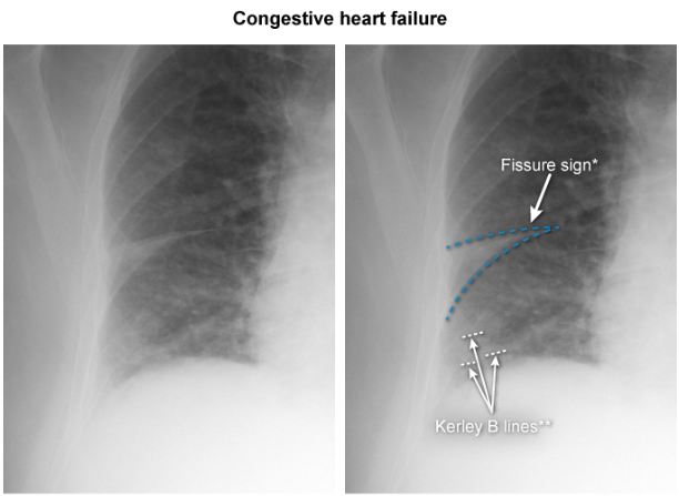
If the LA pressure exceeds 25-30 mmHg, alveolar pulmonary edema may develop, with opacification of the air spaces.
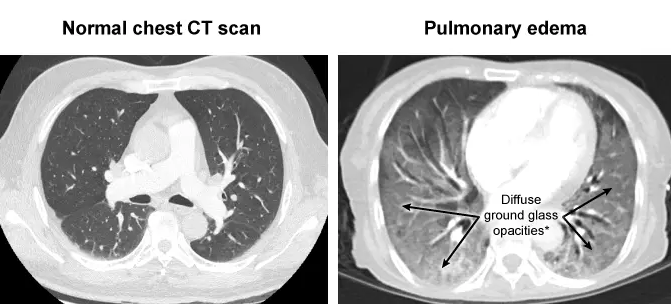
A chest CT may reveal congested lungs with diffuse ground glass opacity and an enlarged cardiac shadow due to heart congestion and enlarged chamber size:
The relationship between LA pressure and chest radiograph findings is modified in patients with chronic heart failure because of enhanced lymphatic drainage, such that higher pressures can be accommodated with fewer radiologic signs.
Assays for BNP correlate well with the degree of LV dysfunction and prognosis. Furthermore, an elevated serum level of BNP can help distinguish heart failure from other causes of dyspnea, such as pulmonary diseases.
The cause of heart failure is often evident from the history, such as a patient who has sustained a large myocardial infarction, or by physical examination, as in a patient with a murmur of valvular heart disease. When the cause is not clear from clinical evaluation, the first step is to determine whether systolic ventricular function is normal or depressed. Of the several noninvasive tests that can help make this determination, echocardiography is especially useful and readily available.
Prognosis
The prognosis of heart failure is poor in the absence of a correctable underlying cause. The 5-year mortality rate following the diagnosis ranges between 45% and 60%, with men having worse outcomes than women.
Patients with severe symptoms (i.e. NYHA class III or IV) are the least well, having a 1-year survival rate of only 40%. The greatest mortality is due to refractory heart failure, but many patients die suddenly, presumably because of associated ventricular arrhythmias.
Acute heart failure
In contrast to chronic heart failure, patients with acute heart failure are those who present with urgent and often life-threatening symptomatology.
Acute heart failure may develop in a previously asymptomatic patient (e.g. from an acute coronary syndrome, severe hypertension, or acute valvular regurgitation), or it may accompany chronic compensated heart failure following a precipitating trigger. Management of acute heart failure typically requires hospitalization and prompt interventions.
The classification acute heart failure and the approach to therapy can be tailored based on the presence or absence of two major findings: volume overload (i.e. “wet” vs. “dry”) as a reflection of LV filling pressures and signs of decreased cardiac output with reduced tissue perfusion (“cold” vs. “warm” extremities). Examples of a “wet” profile, indicative of volume overload, include pulmonary rales, jugular venous distension, and edema of the lower extremities.
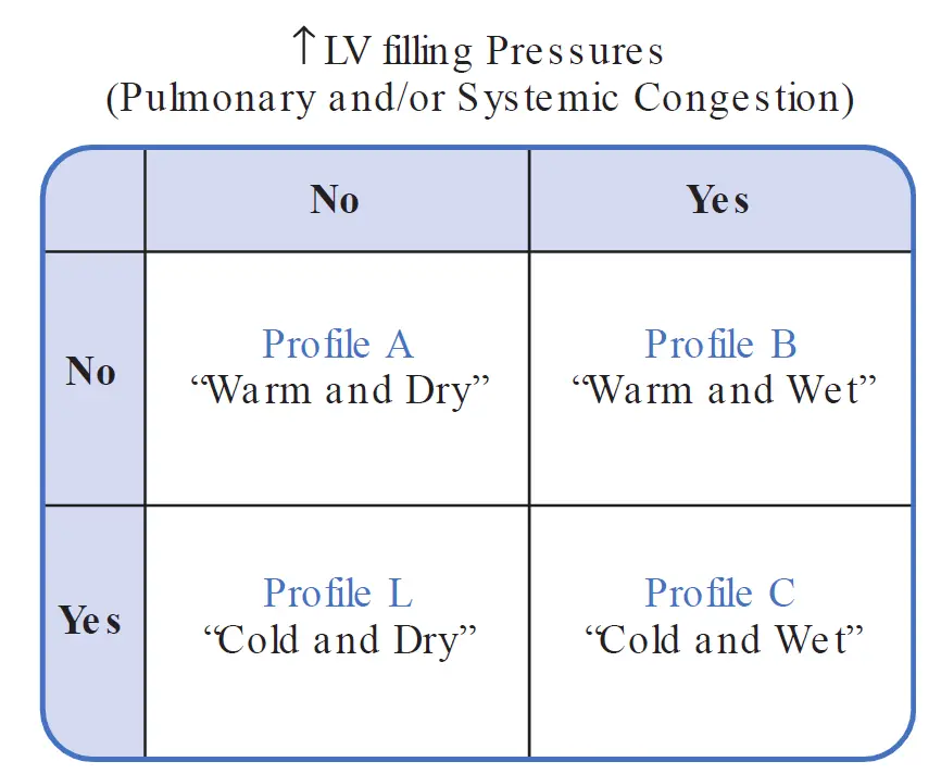
Profile A indicates normal hemodynamics. Cardiopulmonary symptoms in such patients would be due to factors other than heart failure, such as parenchymal lung disease or transient myocardial ischemia.
Profiles B and C are typical of patients with acute pulmonary edema. Profile C is typically more serious than Profile B; in addition to congestive findings, impaired forward cardiac output results in systemic vasoconstriction (e.g. activation of sympathetic nervous system) and therefore “cold” extremities.
Patients with Profile C have a worse prognosis than those with Profile B, who in turn have poorer outcomes than those with Profile A.
Patients with Profile L do not represent an extension of this continuum. Rather, they display “cold” extremities due to low output but no signs of vascular congestion. This profile may arise in patients who are volume deplete, or those with very limited cardiac reserve in the absence of volume overload (e.g. a patient with a dilated left ventricle and mitral regurgitation who becomes short of breath with activity because of the inability to generate adequate forward cardiac output).
Identification of the patient’s profile guides therapeutic interventions. For example, a patient with Profile B would require therapy for pulmonary edema, and those with Profile C may additionally require IV inotropic medications to strengthen cardiac output. Patients with Profile L may require volume expansion. The presence of profile A would prompt a search for contributions to the patient’s symptoms other than heart failure.
Acute Pulmonary Edema
A common manifestation of acute left-sided heart failure is cardiogenic pulmonary edema, in which elevated capillary hydrostatic pressure causes rapid accumulation of fluid within the interstitium and alveolar spaces of the lung. In the presence of normal plasma oncotic pressure, pulmonary edema develops when the pulmonary capillary wedge pressure (reflecting LV diastolic pressure) exceeds approximately 25 mmHg.
This condition is frequently accompanied by hypoxemia because of intra-pulmonary shunting blood. Like other manifestations of acute heart failure, pulmonary edema may appear suddenly in a previously asymptomatic person or in a patient with chronic compensated congestive heart failure following a precipitating event. Acute pulmonary edema is a horrifying experience for the patient, resulting in severe dyspnea and anxiety while struggling to breathe.
On examination, the patient is tachycardic and may demonstrate cold, clammy skin owing to peripheral vasoconstriction in response to increased sympathetic outflow (i.e. Profile C). Tachypnea and coughing of “frothy” sputum represent transudation of fluid into the alveoli. Rales are present initially at the bases and later throughout the lung fields, sometimes accompanied by wheezing because of edema within the conductance airways.
Pulmonary edema is a life-threatening emergency that requires immediate improvement of systemic oxygenation and elimination of the underlying cause. The patient should be seated upright to permit pooling of blood within the systemic veins of the lower body, reducing venous return to the heart. Supplemental oxygen is provided by a face mask. Morphine sulfate is administered intravenously to reduce anxiety and also acts as a venous dilator to facilitate pooling of blood peripherally. A rapidly acting diuretic, such as IV furosemide, is administered to further reduce LV preload and pulmonary capillary hydrostatic pressure. Other means of reducing preload include administration of nitrates (often intravenously). Intravenous inotropic drugs (e.g. dopamine) may increase forward cardiac output and are used primarily in patients with Profile C.
During resolution of the pulmonary congestion and hypoxemia, attention should be directed at identifying and treating the underlying precipitating cause.
An easy-to-remember mnemonic for the principal components of management of acute pulmonary edema is the alphabetic sequence LMNOP:
- Lasix (trade name for furosemide)
- Morphine
- Nitrates
- Oxygen
- Position (sit upright)
Treatment
There are five main goals of therapy in patients with chronic heart failure and a reduced EF:
-
Identification and correction of the underlying condition causing heart failure. In some patients, this may require surgical repair or replacement of dysfunctional valves, coronary artery revascularization, aggressive treatment of hypertension, or cessation of alcohol consumption.
-
Elimination of the acute precipitating cause of symptoms in a patient with heart failure who was previously in a compensated state. This may include, for example, treating acute infections or arrhythmias, removing sources of excessive salt intake, or eliminating drugs that can aggravate symptomatology (e.g. certain calcium channel blockers, which have a negative inotropic effect, or nonsteroidal anti-inflammatory drugs, which can contribute to volume retention).
-
Management of heart failure symptoms. This includes treatment of pulmonary and systemic vascular congestion and measures to increase forward cardiac output and perfusion of vital organs through the use of vasodilators and positive inotropic drugs.
-
Modulation of the neurohormonal response to prevent adverse ventricular remodeling in order to slow the progression of LV dysfunction.
-
Prolongation of long-term survival.
Diuretics
By promoting the elimination of sodium and water through the kidney, diuretics reduce intravascular volume and thus ameliorate the signs of (systemic or pulmonary) congestion. The intent is to reduce the EDP (and therefore hydrostatic forces contributing to pulmonary congestion) without a significant fall in stroke volume.
The judicious use of diuretics does not significantly reduce stroke volume and cardiac output in this setting, because the failing ventricle is operating on the “flat” portion of a depressed Frank–Starling curve. However, overly vigorous diuresis can lower LV filling pressures into the steep portion of the ventricular performance curve, resulting in an undesired fall in cardiac output. Thus, diuretics should be used only if there is evidence of pulmonary congestion or peripheral edema.
Loop diuretics (e.g. furosemide, torsemide, and bumetanide) act primarily at the renal loop of Henle, and are the most potent diuretics in heart failure.
Thiazide diuretics (e.g. hydrochlorothiazide and metolazone) are useful but are less effective in the setting of decreased renal perfusion, which often accompanies heart failure.
The most important side effects in heart failure patients include overly vigorous diuresis resulting in a fall in cardiac output and electrolyte disturbances (particularly hypokalemia and hypomagnesemia), which may contribute to arrhythmias. In patients with acute heart failure exacerbations, diuretics should be administered intravenously because venous congestion can limit the absorption of oral diuretics from the gut.
Vasodilators
One of the most important cardiac advances in the late twentieth century was the introduction of vasodilator therapy or the treatment of heart failure, particularly ACE inhibitors. As described earlier, neurohormonal compensatory mechanisms in heart failure often lead to excessive vasoconstriction, volume retention, and ventricular remodeling, with progressive deterioration of cardiac function. Vasodilators help to reverse these adverse consequences. Moreover, multiple studies have shown that certain vasodilator regimens significantly extend survival in patients with heart failure.
Venous vasodilators (e.g. nitrates) increase venous capacitance and thereby decrease venous return to the heart and left ventricular preload. Consequently, LV diastolic pressures fall and the pulmonary capillary hydrostatic pressure declines, similar to the hemodynamic effects of diuretics. As a result, pulmonary congestion improves, and as long as the heart failure patient is on the relatively “flat” part of the depressed Frank–Starling curve, the cardiac output does not fall despite the reduction in ventricular filling pressure. However, venous vasodilatation in a patient who is operating on the steeper part of the curve may result in an undesired fall in stroke volume, cardiac output, and blood pressure.
Pure arteriolar vasodilators (e.g. hydralazine) reduce systemic vascular resistance and therefore LV afterload, which in turn permits increased ventricular muscle fiber shortening during systole, increasing stroke volume. Although an arterial vasodilator might be expected to reduce blood pressure—an undesired effect in patients with heart failure who may already be hypotensive, this generally does not happen. As resistance is reduced by arteriolar vasodilatation, a concurrent rise in cardiac output usually occurs, such that blood pressure remains constant or decreases only mildly.
Some groups of drugs result in vasodilatation of both the venous and arteriolar pressures (“balanced” vasodilators). Of these, the most important are ACE inhibitors. These interrupt the production of angiotensin II and reduces the levels of aldosterone, facilitating salt and water elimination, resulting in reduced intravascular volume and improvement of congestion symptoms. ACE inhibitors also increase circulating levels of bradykinin, which is a vasodilator that is though to be helpful in heart failure.
As a result of these effects, ACE inhibitors limit maladaptive ventricular remodeling in patients with chronic heart failure and following acute myocardial infarction. Supporting the effectiveness of ACE inhibitors, many large clinical trials have shown that these drugs reduce heart failure symptoms, reduce the need for hospitalization, and most importantly, extend survival in patients with heart failure with reduced EF. Thus, ACE inhibitors are standard first-line chronic therapy for patients with LV systolic dysfunction.
The RAAS can also be therapeutically inhibited by angiotensin II receptor blockers (ARBs). Since angiotensin II can be formed by pathways other than ACE, ARBs provide a more complete inhibition of the system than ACE inhibitors, through blockade of the actual AII receptor. Conversely, ARBs do not cause the potentially beneficial rise in serum bradykinin. The net result is that the hemodynamic effects and mortality benefit of ARBs in heart failure are similar to those of ACE inhibitors. Thus, they are prescribed to heart failure patients mainly when ACE inhibitors are not tolerated (e.g. because of bradykinin-mediated side effects such as cough or angioedema).
Chronic therapy using the combination of the venous dilator isosorbide dinitrate plus the arteriolar dilator hydralazine has also been shown to improve survival in patients with moderate symptoms of heart failure. However, when the ACE inhibitor enalapril was compared with the hydralazine–isosorbide dinitrate (H-ISDN) combination, the ACE inhibitor was shown to produce the greater improvement in survival. Thus, H-ISDN is generally substituted when a patient cannot tolerate ACE inhibitor or ARB therapy (e.g. because of renal insufficiency or hyperkalemia).
Note
The African American Heart Failure trial demonstrated that the addition of H-ISDN to standard heart failure therapy in black patients with heart failure further improved functional status and survival.
Nesiritide (human recombinant BNP) is an intravenous vasodilator drug available for hospitalized patients with decompensated heart failure. It causes rapid and potent vasodilatation, reduces elevated intracardiac pressures, and increases forward cardiac output. However, it is an expensive drug that does not improve outcomes and may worsen renal function, so its use should be restricted to patients who have not responded to, or cannot tolerate other intravenous vasodilators, such as intravenous nitroglycerin or nitroprusside.
Positive Inotropic Drugs
Inotropic drugs include β-adrenergic agonists, digitalis, and phosphodiesterase type 3 inhibitors. By increasing the availability of intracellular calcium, each of these drug groups enhances the force of ventricular contraction and thus shits the Frank–Starling curve in an upward direction. As a result, stroke volume and cardiac output are increased at any given ventricular EDV.
These agents may be useful in treating patients with systolic dysfunction, but typically not those with heart failure with preserved EF.
β-Adrenergic agonists (e.g. dobutamine and dopamine) are administered intravenously for temporary hemodynamic support in acutely ill hospitalized patients. Their long-term use is limited by the lack of an oral form and by the development of drug tolerance possibly owing to down-regulation of myocardial adrenergic receptors.
Likewise, the role of phosphodiesterase 3 inhibitors (e.g. milrinone) is limited to the intravenous treatment of congestive heart failure in acutely ill patients. Despite the initial promise of effectiveness of oral phosphodiesterase 3 inhibitors, studies thus far actually demonstrate reduced survival among patients receiving this form of treatment.
One of the oldest forms of inotropic therapy is digitalis, which can be administered intravenously or orally. Digitalis enhances contractility, improves symptoms, and increases cardiac output in patients with systolic heart failure. Digitalis also increases the sensitivity of the baroreceptors, so that the compensatory sympathetic drive in heart failure is reduced, reducing left ventricular afterload. It also slows down AV nodal conduction, reducing the heart rate, which can be beneficial in patients with congestive heart failure who have concurrent atrial fibrillation.
Although digitalis can improve the symptoms and reduce the rate of hospitalizations in heart failure, it has not been shown to improve long-term survival. Thus, its use is limited to patients who remain symptomatic despite other standard therapies or to help slow the ventricular rate if atrial fibrillation is present. Digitalis is not useful in the treatment of heart failure with preserved EF because it does not improve ventricular relaxation properties.
β-Blockers
Historically, β-blockers were thought to be contraindicated in patients with systolic dysfunction because their negative inotropic effect would be expected to worsen the symptoms. However, clinical trials have actually shown that long-term β-blocker therapy has important benefits in patients with stable chronic heart failure with reduced EF, including increasing cardiac output, reducing the need for hospitalizations, and improving survival.
The explanation for these desired effects remains conjectural but may relate to the drugs’ effects on reducing heart rate and blunting chronic sympathetic activation or to their anti-ischemic properties.
The 3 β-blockers that have been shown to be beneficial in randomized clinical trials of heart failure include carvedilol (nonselective β-blocker with weak α-blocking properties) and the β1-selective agents metoprolol succinate and bisoprolol. These drugs are well tolerated in stable patients. Nonetheless, β-blockers should always be used cautiously in heart failure to prevent acute deterioration related to their negative inotropic effect. Regimens should start at low dosage and increase gradually.
Aldosterone Antagonist Therapy
There is evidence that chronic excess of aldosterone in heart failure contributes to cardiac fibrosis and adverse ventricular remodeling. Antagonists of this hormone (which have been used historically as mild diuretics) have shown clinical benefits in heart failure.
For example, in a clinical trial of patients with advanced heart failure (i.e. NYHA Class III to IV) who were already taking an ACE inhibitor and diuretics, the aldosterone receptor antagonist spironolactone substantially reduced mortality rates and improved heart failure symptoms. Eplerenone, a more specific aldosterone receptor inhibitor, has been shown to improve survival of patients with congestive heart failure after acute myocardial infarction as well as patients with more mild forms of chronic heart failure (i.e. NYHA Class II to III).
Although aldosterone antagonists are well tolerated, serum potassium level must be monitored to prevent hyperkalemia, especially if there is renal impairment or concomitant ACE inhibitor therapy.
Additional therapy
Intraventricular conduction abnormalities with widened QRS complexes are common in patients with advanced heart failure. Such abnormalities can contribute to the cardiac symptoms because of the uncoordinated pattern of right and left ventricular contraction. Advanced pacemakers have been developed that stimulate both ventricles simultaneously, thus resynchronizing the contractile effort. This technique of biventricular pacing, also termed cardiac resynchronization therapy (CRT) , has been shown to improve left ventricular systolic function, improve exercise capacity, and reduce the frequency of heart failure exacerbations and mortality.
Thus, CRT is appropriate for selected patients with advanced systolic dysfunction (LV EF ), a prolonged QRS duration ( ms) and continued symptoms of heart failure despite appropriate pharmacologic therapies. Since patients who receive CRT are typically also candidates for an implantable cardioverter-defibrillator, modern devices combine both functions in a single, small implantable unit.
A patient with severe LV dysfunction whose condition remains refractory to maximal medical management may be a candidate for cardiac transplantation. However, only approximately 4,000 transplants are performed worldwide each year because of a shortage of donor hearts, much fewer than the number of patients with refractory heart failure symptoms. For certain patients who are too ill to wait for a heart donor, or who are not eligible for a transplant, alternative mechanical therapies can be used in select patients. Ventricular assist devices (VADs) and implantable total artificial hearts can be used to support cardiac pump function in such patients. Recent technological advances in continuous-flow left-sided VADs have resulted in 1-year survival rates greater than 70%, compared to less than 25% survival rates in similar groups of advanced heart failure patients treated with medical therapy alone.
See also
References
Additional Reading
- Braunwald E. Heart failure. JACC: Heart Failure. 2013;1:1–20.
- Hsich EM, Pina IL. Heart failure in women. J Am Coll Cardiol. 2009;54:491–498.
- Maeder MT, Kaye DM. Heart failure with normal left ventricular ejection fraction. J Am Coll Cardiol. 2009;53:905–918.
- Maron BA, Leopold JA. Aldosterone receptor antagonists: effective but often forgotten. Circulation. 2010;121:934–939.
- McMurray JJV. Systolic heart failure. N Engl J Med. 2010;362:228–238.
- Stewart G, Givertz M. Mechanical circulatory support for advanced heart failure: patients and technology in evolution.Circulation. 2012;125:1304–1315.
- Triposkiadis F, Karayannis G, Giamouzis G, et al. The sympathetic nervous system in heart failure: physiology, pathophysiology, and clinical implications. J Am Coll Cardiol. 2009;54:1747–1762.
- Yancy C, Jessup M, Bozkurt B, et al. 2013 ACCF/ AHA guideline for the management of heart failure: executive summary a report of the American College of Cardiology Foundation/American Heart Association Task Force on Practice Guidelines. Circulation. 2013;128:1810–1852