Part 11: Hypertension
Mon Aug 03 2026
By B. Hassan
Hypertension affects a large portion of the population, and will increase with the aging population; data from the Framingham Heart Study indicate that 90% of people over age 55 will develop hypertension during their lifetimes.
Hypertension represents a great public health concern because it is a major risk factor for coronary artery disease, stroke, heart failure, renal disease, and peripheral vascular disease. Surprisingly, two thirds of hypertensive patients are either unaware of their condition or are not treated adequately to minimize cardiovascular risk. Moreover, because elevated BP is usually asymptomatic until an acute cardiovascular event strikes, screening for hypertension is a critical aspect of preventive medicine.
Hypertension is also a scientific problem of unexpected complexity. In approximately 90% of affected patients, the cause of the elevated BP is unknown, a condition termed primary or essential hypertension (EH).
High BP attributed to a definable cause is termed secondary hypertension. Although far less common than EH, conditions that cause secondary hypertension are important because many are amenable to permanent cure. Many of the conditions now understood to cause secondary hypertension were once unknown, and affected patients were considered to have EH. As more is learned about the pathophysiology of high BP, fewer cases will likely be considered to have the essential type.
BP varies widely in the population and tend to increase with age. The risk of a vascular complication increases progressively and linearly with higher BP values, so the exact cutoff points to define stages of hypertension are somewhat arbitrary. The currently accepted criteria for defining hypertension are as follows:
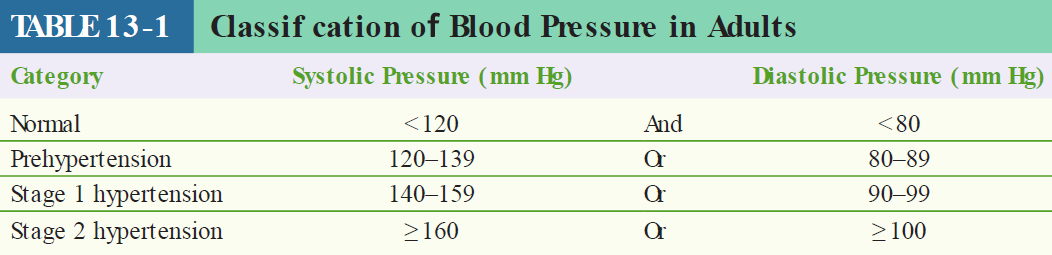
By this classification, a diastolic pressure consistently mmHg or a systolic pressure mmHg establishes the diagnosis of hypertension. Those with prehypertension have an increased risk of developing definite hypertension over time. Although the emphasis has historically been on the level of diastolic pressure, more recent evidence suggests that systolic pressure more accurately predicts cardiovascular complications.
Physiological blood pressure regulation
BP is the product of cardiac output (CO) and total peripheral resistance (TPR):
And CO is the product of the stroke volume (SV) and heart rate (HR):
As described in part 8, SV is determined by cardiac contractility; venous return to the heart (preload); and the resistance the left ventricle must overcome to eject blood into the aorta (afterload).
At least four main systems are directly responsible for BP regulation: the heart, which supplies the pumping pressure; the blood vessel tone, which largely determines systemic resistance; the kidney, which regulates intravascular volume; and hormones, which modulate the functions of the other three systems.
The renal component of BP regulation deserves special mention. No matter how high the CO or TPR, renal excretion has the capacity to completely return BP to normal by reducing intravascular volume. Thus, the development of chronic hypertension requires renal participation. Transplantation studies have confirmed this point: the implantation of a kidney from a normotensive person into a hypertensive one typically improves the BP. Similarly, transplantation of a kidney from a hypertensive rat into a previously normotensive one usually leads to hypertension.
In the presence of normally functioning kidneys, an increase in BP leads to increased urine volume and sodium excretion, which returns the BP to normal. This process, known as pressure natriuresis, is blunted in the kidneys of hypertensive patients; thus, higher pressures are required to excrete a given sodium and water load. Current evidence suggests at least two possible reasons for this blunted response. First, microvascular injury in the kidneys of hypertensive patients impairs sodium excretion. Second, the defect may lie with hormonal factors critical to appropriate renal reaction to the sodium and intravascular volume environment (e.g. the renin–angiotensin system).
BP reflexes
The cardiovascular system has various feedback mechanisms that continuously monitor BP and then respond rapidly to those changes. One such mechanism is the baroreceptor reflex, which is mediated by receptors in the walls of the aortic arch and the carotid sinuses. The baroreceptors monitor changes in pressure by sensing the stretch of the arteries. The higher the BP, the more the baroreceptors are stretched and the greater the impulse transmission rate to the brain. Negative feedback signals are then sent back to the circulation via the autonomic nervous system, causing the BP to fall back to baseline.
Signals from the carotid sinus receptors are carried by the glossopharyngeal nerve (cranial nerve IX), whereas those from the aortic arch receptors are carried by the vagus nerve (cranial nerve X). These nerves converge at the tractus solitarius in the medulla, where the baroreceptor impulses inhibit sympathetic nervous system outflow and excite parasympathetic effects.
The net result is vasodilation (decreasing TPR) and a reduction in CO (because of a lower HR and reduced contractility). Each of these effects tend to lower arterial pressure back towards baseline. Conversely, when a fall in systemic pressure is sensed by the baroreceptors, fewer impulses are transmitted to the medulla, leading to a reflexive increase in BP.
The main effect of the baroreceptor mechanism is to modulate moment-by-moment variations in systemic BP. However, the reflex is not involved in chronic regulation of BP and does not prevent the development of chronic hypertension. This is because of the high adaptability of the baroreceptors such that after a day or two of exposure to higher-than-baseline BP, the baroreceptor-firing rate slows back to its control value, and a new set point is established.
Essential hypertension (EH)
Approximately 90% of hypertensive patients have BPs that are elevated for no readily definable reason. It is a diagnosis of exclusion, and clinicians have to consider other causes of secondary hypertension before EH is diagnosed.
EH is more of a description than a diagnosis, indicating that a patient has a high BP for which no cause has been found. In all likelihood, different underlying defects are responsible for the elevated pressure in different subpopulations of patients. The picture that emerges from many research is that EH likely results from multiple defects of BP regulation that interact with environmental stressors. The regulatory defects may be acquired or genetically determined and may be independent of one another.
Genetics and Epidemiology
Strong support for the role of heredity in EH is evident in the higher rate of elevated BP among first-degree relatives of hypertensive patients than in the general population. Further, concordance between identical twins is high and significantly greater than it is that between dizygotic twins. However, no singular, consistent genetic marker for hypertension has been identified, and it seems likely that EH is a complex polygenic disorder.
While autosomal dominant contributors to elevated BP have been discovered, these are rare and are thought to represent only a small fraction of patients. With respect to genes that affect hypertension in a polygenic way, genes regulating the renin–angiotensin–aldosterone system have been most thoroughly studied because of the central role of this system in affecting BP. Within this group, certain polymorphisms in the genes for angiotensinogen, angiotensin-converting enzyme (ACE), the angiotensin type-1 receptor, and aldosterone synthase confer a small increase in the risk of developing hypertension. Additionally, polymorphisms in the gene for alpha-adducin, a cytoskeletal protein, may be involved in a subgroup of EH patients, possibly by increasing renal tubular sodium absorption.
Finally, significant associations exist among hypertension and obesity, insulin resistance, and diabetes. These conditions are all characterized by similar complex inheritance patterns, some of which may overlap with the genetic underpinnings of hypertension.
As genetics alone cannot explain the complete basis of hypertension, it stands that the environment also plays a role. Indeed, hypertension has been linked to low socioeconomic status, certain dietary and exercise patterns, poor access to health care, and comorbid medical conditions such as obesity, diabetes, and kidney disease. Thus, the heritable traits described above most likely predispose to hypertension after exposure to certain environmental triggers.
Systemic Abnormalities
Multiple defects in BP regulation have been found in EH patients and their relatives. By themselves, or in conjunction with one another, these abnormalities may contribute to chronic BP elevation.
The heart can contribute to a high CO-based hypertension owing to sympathetic overactivity. For example, when tested under psychologically stressful conditions, hypertensive patients often develop excessive HR acceleration compared with control subjects, suggesting an excessive sympathetic response.
Blood vessels may contribute to peripheral vascular resistance–based hypertension by constricting in response to increased sympathetic activity; abnormal regulation of vascular tone by local factors, including nitric oxide, endothelin, and natriuretic actors; or ion channel defects in contractile vascular smooth muscle.
The kidney can induce volume-based hypertension by retaining excessive sodium and water as a result of failure to regulate renal blood flow appropriately; ion channel defects, which directly cause sodium retention; or inappropriate hormonal regulation. For example, renin levels in EH patients (compared with those in normotensive persons) are subnormal in 25%, normal in approximately 60%, and high in 10% to 15%. Because renin secretion should be suppressed by high BP, even “normal” levels are inappropriate in hypertensives.
Metabolic syndrome
Recent research suggests that insulin may play a role in the development of EH. In many people with hypertension (especially those who are obese or have type 2 diabetes,) insulin resistance is usually present. This causes insulin levels to be chronically elevated, which may contribute to hypertension via increased sympathetic activation or by stimulation of vascular smooth muscle hypertrophy.
Smooth muscle hypertrophy may be caused by a direct mitogenic effect of insulin or through enhanced sensitivity to platelet-derived growth factor.
Obesity itself has been directly associated with hypertension. Possible explanations include the release of angiotensinogen from adipocytes; increased blood volume related to increased body mass; and increased blood viscosity caused by adipocyte release of profibrinogen and plasminogen activator inhibitor 1.
Natural course of EH
EH characteristically arises after young adulthood. Its prevalence increases with age, and more than 60% of Americans older than 60 years are hypertensive.
In addition, the hemodynamic characteristics of BP elevation in EH tend to change over time. The systolic pressure increases throughout adult life, while the diastolic pressure rises until about the age of 50 and then declines slightly thereafter. Accordingly, diastolic hypertension is more common in young people, while a substantial number of hypertensive patients over 50 have isolated systolic hypertension with normal diastolic values.
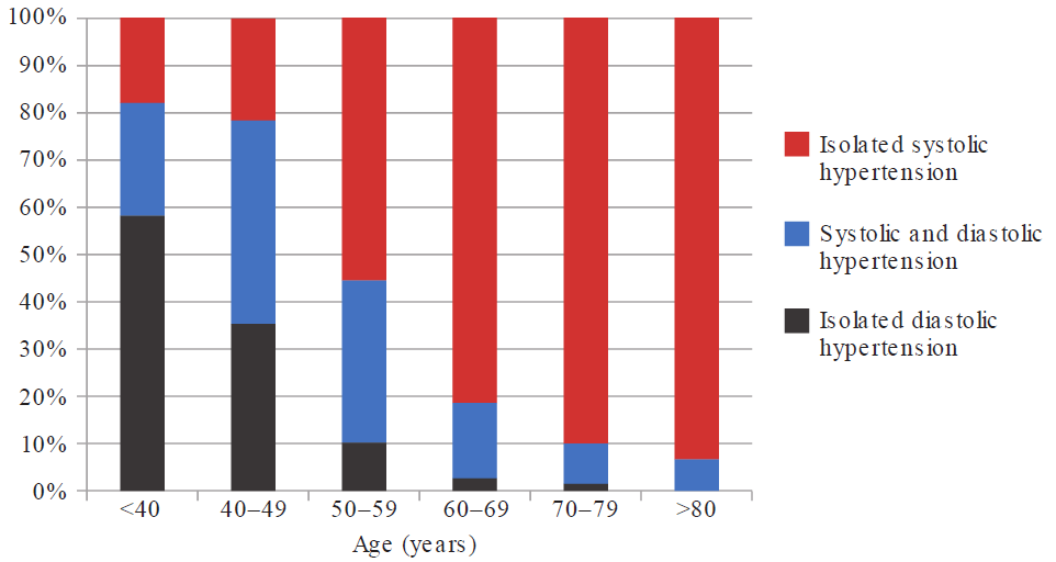
In younger patients with hypertension, elevated BP tends to be driven by high CO in the setting of relatively normal peripheral vascular resistance, termed the hyperkinetic phase of EH. With advancing age, however, the effect of CO declines, perhaps because of the development of left ventricular hypertrophy and its consequent reduced diastolic filling (which in turn reduces SV and CO). Conversely, vascular resistance increases with age due to medial hypertrophy as the vessels adapt to the prolonged pressure stress. Thus, younger hypertensive patients often display increased CO as the principal abnormality, and older patients tend to have elevated TPR as the major finding.
Secondary hypertension
Although EH is more common, a defined cause of hypertension may be found in a small percentage of patients. Identification of such cases is important because the underlying conditions may require therapy different from that of EH, and they are often curable. Moreover, if secondary hypertension is left uncontrolled, adaptive cardiovascular changes may develop analogous to those of long-standing EH that could cause the elevated pressures to persist even after the underlying cause is corrected.
Although secondary forms should be considered in the workup for all patients with hypertension, there are clues that a given patient may have one of the correctable conditions:
-
If a patient develops hypertension before age 20 or after age 50, secondary hypertension is more likely.
-
Secondary hypertension often causes BP to rise dramatically, whereas most EH patients usually have mild-to-moderate hypertension.
-
Secondary forms of hypertension often present abruptly in a patient who was previously normotensive, rather than gradually progressing over years as is in EH.
-
EH patients often have hypertensive first-degree relatives, whereas secondary hypertension more commonly occurs sporadically.
The process that induces hypertension may give rise to other characteristic abnormalities, identified by the history and physical examination. For example, a renal artery bruit may be heard on abdominal examination in a patient with renal artery stenosis. Excessive weight loss may be an indicator of pheochromocytoma, whereas weight gain may point to the presence of Cushing syndrome.
The history should also include an assessment of lifestyle behaviors that may contribute to hypertension such as smoking, and the patient’s medications should be reviewed because certain drugs may elevate BP.
Obstructive sleep apnea is commonly associated with hypertension and should be considered particularly in patients who snore and have a history of hypertension refractory to medications.
Laboratory tests commonly performed for the evaluation of hypertensive patients, including general screening for secondary causes are:
-
Urinalysis, serum creatinine, and blood urea nitrogen
-
Serum potassium level (abnormally low in renovascular hypertension or aldosteronism)
-
Blood glucose level (elevated in diabetes, which is strongly associated with hypertension and renal disease)
-
Serum cholesterol, HDL cholesterol, and triglyceride levels, as part of the global vascular risk screen
-
An electrocardiogram (for evidence of LVH caused by chronic hypertension).
If no abnormalities are found that suggest a secondary cause for hypertension, the patient is presumed to have EH and treated accordingly. If, however, the patient’s BP continues to be elevated despite standard treatments, then more detailed diagnostic testing may be undertaken to search for specific secondary causes.
Exogenous Causes
Several medications can elevate BP. For example, oral contraceptives may cause secondary hypertension in some women. The mechanism is likely related to increased activity of the renin–angiotensin system. Estrogens increase the hepatic synthesis of angiotensinogen, leading to greater production of angiotensin II.
Other medications that can raise BP include corticosteroids, cyclosporine (antirejection drug used in organ transplants), erythropoietin (a hormone that increases bone marrow RBC formation and elevates BP by increasing blood viscosity and reversing local hypoxic vasodilatation), and sympathomimetic drugs. Nonsteroidal anti-inflammatory drugs can contribute to hypertension through dose-related increase in renal sodium and water retention.
Two other substances that may contribute to hypertension are ethanol and cocaine, which are both associated with increased sympathetic nervous system activity.
Renal Causes
Renal disease contributes to two important endogenous causes of secondary hypertension: renal parenchymal disease, accounting for 2% to 4% of hypertensive patients, and renal arterial stenosis, which accounts for approximately 1%.
Parenchymal kidney damage can result from diverse pathologies. The major mechanism by which injury leads to elevated BP is through increased intravascular volume as damaged nephrons are unable to excrete normal amounts of sodium and water, leading to a rise in intravascular volume. If renal function is only mildly impaired, BP may stabilize at a level at which the higher systemic pressure (and therefore renal perfusion pressure) enables sodium excretion to balance sodium intake. Conversely, if a patient has end-stage renal failure, glomerular filtration rate may be greatly decreased that the kidneys simply cannot excrete sufficient volume, and malignant-range BP may follow.
Stenosis of one or both renal arteries leads to hypertension. Although emboli, vasculitis, and external compression of the renal arteries can be responsible, the two most common causes are atherosclerosis and fibromuscular dysplasia. Atherosclerotic lesions account for about two thirds of cases with renal artery stenosis and occurs mainly in elderly men. In contrast, fibromuscular lesions consist of discrete regions of fibrous or muscular proliferation and characteristically occurs in young women. The elevated BP arises from reduced renal blood flow to the affected kidney, which responds to the lower perfusion pressure by secreting renin, activating the renin-angiotensin aldosterone system.
The diagnosis of renal artery stenosis is suggested by an abdominal bruit, which can be found in 40% to 60% of patients, or by the presence of unexplained hypokalemia (owing to excessive renal excretion of potassium as a result of an elevated aldosterone level). The diagnosis can be confirmed by Doppler ultrasonography, CT angiography, or MRI of the renal arteries.
Therapy for renal artery stenosis with antihypertensive drugs is often effective, particularly when an ACE inhibitor or angiotensin receptor blocker is included. These antagonize the effects of elevated circulating renin by impeding the formation or action of angiotensin II. However, these should be avoided in patients with bilateral renal artery stenosis as the inhibition of angiotensin II may excessively reduce intraglomerular pressure and filtration, and worsen renal function in patients with bilateral disease who already have compromised perfusion to both kidneys. In select patients, (e.g. those with recent onset hypertension due to renal artery stenosis, those whose BP remains elevated despite medical therapy, or those with progressive renal insufficiency due to renal artery stenosis) percutaneous catheter interventions or surgical reconstruction of the stenotic vessel may be more effective than continued medical antihypertensive therapy alone.
Coarctation of the Aorta
Coarctation is an infrequent congenital narrowing of the aorta typically just distal to the origin of the left subclavian artery. As a result of the relative obstruction, the BP in the aortic arch, head, and arms is higher than that in the descending aorta and its branches and in the lower extremities. Sometimes the coarctation involves the origin of the left subclavian artery, causing lower pressure in the left arm compared with the right arm.
Hypertension in this condition arises by two mechanisms. First, reduced blood flow to the kidneys stimulates the renin–angiotensin aldosterone system. Second, high pressures proximal to the coarctation stiffen the aortic arch through accelerated atherosclerosis, blunting the normal baroreceptor reflex.
Clinical clues to the presence of coarctation include symptoms of inadequate blood flow to the legs or left arm, such as claudication or fatigue, or the finding of weakened or absent femoral pulses. A mid-systolic murmur associated with the stenotic segment of the aorta may be auscultated, especially over the back, between the scapulae.
Chest radiography may show indentation of the aorta at the level of the coarctation. It may also demonstrate a notched appearance of the ribs secondary to the enlargement of collateral intercostal arteries, which shunt blood around the aortic narrowing.
Treatment options include angioplasty or surgery to correct the stenosis. However, hypertension may not disappear completely after mechanical correction, in part because of persistent desensitization of the arterial baroreceptors.
Pheochromocytoma
Pheochromocytomas are catecholamine-secreting tumors of neuroendocrine cells (usually in the adrenal medulla) that account for approximately 0.2% of cases of hypertension.
It releases epinephrine and norepinephrine, resulting in intermittent or chronic vasoconstriction, tachycardia, and other sympathetic-mediated effects. A characteristic presentation consists of paroxysmal rises in BP accompanied by “autonomic attacks” caused by the increased catecholamine levels: severe throbbing headaches, profuse sweating, palpitations, and tachycardia. Although some patients are actually normotensive between attacks, most have sustained hypertension.
Determination of plasma catecholamine levels, or urine catecholamines and their metabolites (e.g. vanillylmandelic acid and metanephrine) are used to identify this condition. Because some pheochromocytomas secrete only episodically, diagnosis may require measurement of catecholamines immediately following an attack.
Pharmacologic therapy of pheochromocytomas includes the combination of an α-blocker (e.g. phenoxybenzamine) and a β-blocker. However, once the tumor is localized by CT, MRI, or angiography, the definitive treatment is surgical resection. For patients with inoperable disease, treatment consists of α- and β-blockade as well as drugs that inhibit catecholamine biosynthesis (e.g. α-methyltyrosine).
Adrenocortical Hormone Excess
Among the hormones produced by the adrenal cortex are mineralocorticoids and glucocorticoids. Excess of either of these can result in hypertension.
Mineralocorticoids, primarily aldosterone, increase blood volume by stimulating reabsorption of sodium into the circulation by the distal portions of the nephron in exchange for potassium excretion into the urine. Primary hyperaldosteronism results either from an adrenal adenoma (termed Conn syndrome) or from bilateral hyperplasia of the adrenal glands.
While once considered rare, recent data suggest that the frequency of primary hyperaldosteronism may be as high as 10% to 15% among hypertensives, depending on the sensitivity of screening, with a substantial majority having the bilateral hyperplasia form. The diagnosis may be suspected by the presence of hypokalemia and is confirmed by the finding of excessive plasma aldosterone and a suppressed renin level. Therapy includes either surgical removal of the responsible adenoma (if present) or medical management with aldosterone receptor antagonists.
Glucocorticoid-remediable aldosteronism (GRA), an uncommon autosomal dominant form of primary aldosteronism resulting from a genetic defect in which aldosterone synthesis abnormally comes under the regulatory control of adrenocorticotropic hormone (ACTH). This condition typically presents as severe hypertension in childhood or young adulthood, as opposed to the more common forms of primary aldosteronism, which are generally diagnosed in the third through sixth decades. Unlike other forms of hypertension, GRA-related BP elevation responds to glucocorticoid therapy, which suppresses ACTH release from the pituitary gland.
Secondary aldosteronism can result from increased angiotensin II production stimulated by rare renin-secreting tumors. More commonly, secondary elevation of aldosterone is a result of increased circulating angiotensin II in women taking oral contraceptives or because of impaired angiotensin II degradation in chronic liver diseases.
Glucocorticoids elevate BP when present in excess, likely via blood volume expansion and stimulated synthesis of components of the renin–angiotensin system. In addition, though mineralocorticoids are more potent activators of mineralocorticoid receptors in the renal tubules, excess glucocorticoids may also activate them. Nearly 80% of patients with Cushing syndrome, a disorder of glucocorticoid excess, have some degree of hypertension. These patients often present with classic “cushingoid” features: a characteristic rounded facial appearance, central obesity, proximal muscle weakness, and hirsutism. The cause of the excess glucocorticoids may be either an ACTH-secreting tumor/adenoma (causing adrenal cortical hyperplasia) or an adrenal cortisol-secreting adenoma. The diagnosis of Cushing syndrome is confirmed by a 24-hour urine collection for the measurement of cortisol, or by a dexamethasone test, which evaluates whether exogenous glucocorticoids can suppress cortisol secretion.
Thyroid Hormone Abnormalities
Approximately one third of hyperthyroid and one fourth of hypothyroid patients have significant hypertension.
Thyroid hormones exert their cardiovascular effects by increasing blood volume and by stimulating tissue metabolism and oxygen demand, with secondary accumulation of metabolites that modulate local vascular tone.
Hypothyroid patients demonstrate predominantly diastolic hypertension and an increase in peripheral vascular resistance. Though the precise mechanism is unclear, the effect appears to be mediated by sympathetic and adrenal activation linked to hypothyroidism.
Consequences of hypertension
In the past, “classic” symptoms of hypertension included headache, epistaxis (nose bleeds), and dizziness. However, the usefulness of these symptoms has been questioned by studies that show that they are found no more frequently among hypertensive patients than in the general population. Other symptoms, such as flushing, sweating, and blurred vision, do seem more common in the hypertensive population. In general, however, most hypertensive patients are asymptomatic and are diagnosed simply by BP measurement during routine physical examinations.
Target organ complications of hypertension reflect the degree of chronic BP elevation. This damage is mainly attributed to vascular damage as a result of the elevated blood pressure causing smooth muscle hypertrophy and endothelial dysfunction, ultimately accelerating the atherosclerotic process. Arteries lined by atherosclerotic plaque may thrombose or may serve as a source of cholesterol emboli that can cause organ infarction. In addition, atherosclerosis of large arteries hinders their elasticity, resulting in systolic pressure spikes that can further damage endothelium or provoke events such as aneurysm rupture.
The major target organs for complications of chronic hypertension are the heart, cerebrovascular system, the vascular system, the kidney, and the retina. Left untreated, approximately 50% of hypertensive patients die of coronary artery disease or congestive heart failure, about 33% succumb to stroke, and 10% to 15% die from complications of renal failure.
Heart
The major cardiac effects of hypertension relate to the increased afterload and accelerated atherosclerosis within the coronary arteries. The high afterload increases wall tension of the left ventricle, which is compensated mainly through concentric hypertrophy (without dilatation), although conditions that elevate BP by virtue of increased circulating volume (e.g. primary aldosteronism) may instead cause eccentric hypertrophy with chamber dilatation.
Left ventricular hypertrophy (LVH) results in increased stiffness of the left ventricle with diastolic dysfunction, manifested by increased LV filling pressures that can result in heart failure with preserved ejection fraction. LVH is one of the strongest predictors of cardiac morbidity in hypertensive patients.
Physical findings of LVH may include a heaving LV impulse on chest palpation, indicative of the increased muscle mass. It is frequently accompanied by an S4 sound as the left atrium contracts into the stiffened left ventricle.
Although LVH initially serves a compensatory role, later in the course of systemic hypertension, the increased LV mass may be insufficient to balance the high wall tension caused by the elevated pressure. As LV contractile capacity deteriorates, findings of systolic dysfunction become evident. Systolic dysfunction is also provoked by the accelerated development of coronary artery disease with resultant periods of myocardial ischemia.
Cerebrovascular System
Hypertension is the major modifiable risk factor for stroke, also termed cerebrovascular accidents (CVAs). Although diastolic pressure is important, it is the systolic pressure that has been most closely linked to CVAs. The presence of isolated systolic hypertension more than doubles a person’s risk for this complication.
Hypertension-induced strokes can be hemorrhagic or, more commonly, atherothrombotic. Hemorrhagic CVAs result from rupture of microaneurysms induced in cerebral parenchymal vessels due to long-standing hypertension. Atherothrombotic (also called thromboembolic) CVAs arise when portions of atherosclerotic plaque within the carotids or major cerebral arteries, or thrombi that form on those plaques, break off and embolize to smaller distal vessels.
Occlusion of small penetrating brain arteries can result in multiple tiny infarcts. As these lesions are absorbed by phagocytic cells, small ( mm diameter) cavities form, termed lacunae. These lacunar infarctions are seen almost exclusively in patients with long-standing hypertension.
In addition, the generalized arterial narrowing found in hypertensive patients reduces collateral flow to ischemic tissue and imposes the need for higher perfusion pressure to maintain adequate tissue flow. This leaves the hypertensive patient vulnerable to cerebral infarcts in areas supplied by the distal ends of arterial branches (“watershed” infarcts) if BP should fall suddenly.
Peripheral vasculature
The accelerated atherosclerosis associated with hypertension may result in plaque formation and narrowing throughout the arterial vasculature. In addition to the coronary arteries, lesions most commonly appear within the aorta and the major arteries that serve the lower extremities, neck, and brain.
Chronic hypertension may lead to the development of aneurysms, particularly of the abdominal aorta, usually located below the level of the renal arteries. This is usually the result of the mechanical stress of the high pressure on an arterial wall already weakened by atherosclerosis. Aneurysms cm in diameter have a very high likelihood of rupture within 2 years if not surgically corrected.
Another life-threatening vascular complication of high BP is aortic dissection as elevated BP accelerates degeneration in the media of the aorta. When the weakened wall is further exposed to high pressure, the intima may tear, allowing blood to dissect into the aortic media and propagate in either its normal direction or in the false lumen formed by the dissection. This may “clip off” and obstruct major branch vessels along the way (including coronary or carotid arteries). The treatment of aortic dissection requires rigorous BP control and urgent surgical repair if the proximal aorta is involved.
Kidney
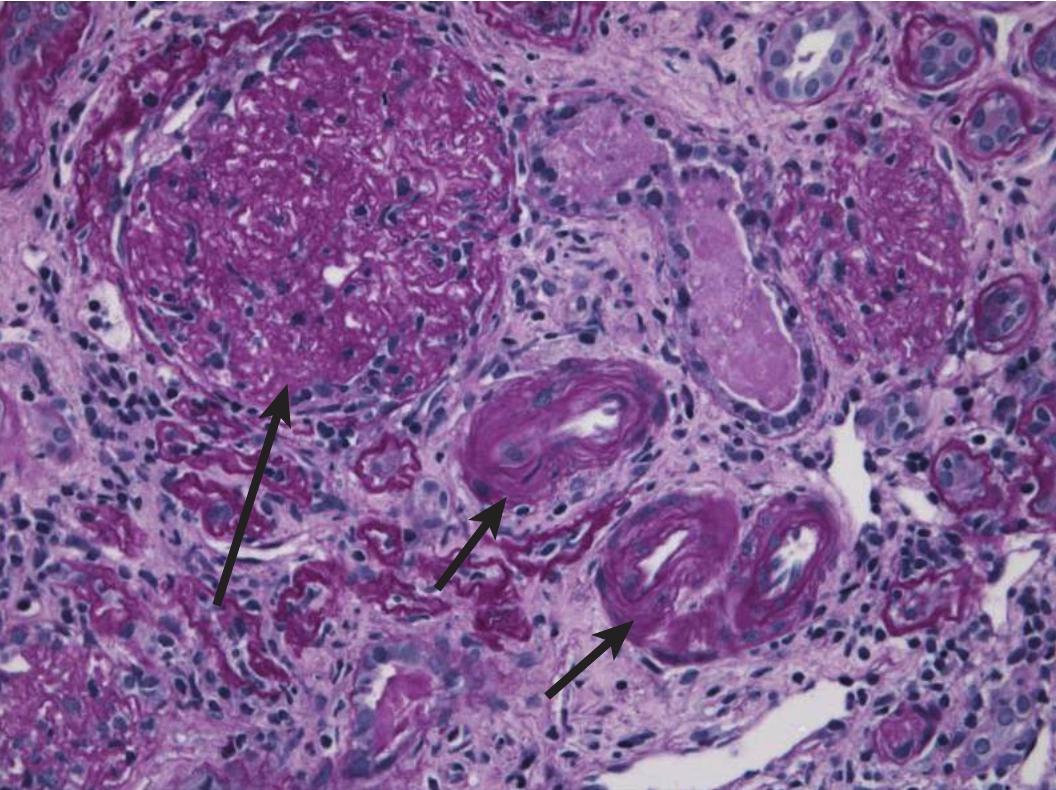
Hypertension-induced kidney disease (nephrosclerosis) is a leading cause of renal failure that results from damage to the organ’s vasculature. Histologically, the vessel walls become thickened with a hyaline infiltrate known as hyaline arteriolosclerosis. Hypertension also induces smooth muscle hypertrophy and necrosis of capillary walls, termed fibrinoid necrosis.
The following image shows nephrosclerosis. The arteriolar walls are thickened by hyaline infiltrate (short arrows). The glomeruli (long arrow) appear sclerosed because of reduced vascular supply.
These changes result in reduced vascular supply and subsequent ischemic atrophy of renal tubules and, to a lesser extent, glomeruli. Because intact nephrons can usually compensate for those damaged by ischemia, mild hypertension rarely leads to renal insufficiency in the absence of other causes of renal damage. However, malignant levels of hypertension can inflict permanent damage leading to chronic renal failure.
One of the consequences of hypertensive renal failure is perpetuation of elevated BP. For example, progressive renal dysfunction compromises the ability of the kidney to regulate blood volume, which contributes further to chronic hypertension.
Retina
High BP induces abnormalities that are collectively termed hypertensive retinopathy. Although vision may be compromised when the damage is extensive, more commonly the changes serve as an asymptomatic clinical marker for the severity of hypertension and its duration. Severe hypertension that is acute in onset may burst small retinal vessels, causing hemorrhages, exudation of plasma lipids, and areas of local infarction.
If ischemia of the optic nerve develops, patients may describe generalized blurred vision. Retinal ischemia caused by hemorrhage leads to more patchy loss of vision. Papilledema, or swelling of the optic disk with blurring of its margins, may arise from high intracranial pressure when the BP reaches malignant levels and cerebrovascular autoregulation begins to fail.
Chronically elevated BP results in a different set of retinal findings. Papilledema is absent, but vasoconstriction results in arterial narrowing, and medial hypertrophy thickens the vessel wall, which “nicks” (indents) crossing veins. With more severe chronic hypertension, arterial sclerosis is evident as an increased reflection of light through the ophthalmoscope (termed “copper” or “silver” wiring). Although these changes are not in themselves of major functional importance, they indicate that the patient has had long-standing, poorly controlled hypertension.
Hypertensive crisis
A hypertensive crisis is a medical emergency characterized by a severe elevation of BP. In the past, this type of elevation was usually a consequence of inadequate BP treatment. Now a hypertensive crisis is more often caused by an acute hemodynamic event (e.g. acute renal disease) superimposed on a chronic hypertensive state. As a result of rapid pathologic changes (fibrinoid necrosis) within the blood vessels and kidneys, a spiraling increase in BP evolves. Further volume expansion and vasoconstriction occur as renal perfusion drops and serum renin and angiotensin levels rise.
Severe BP elevation results in increased intracranial pressure, and patients may present with hypertensive encephalopathy manifested by headache, blurred vision, confusion, somnolence, and sometimes coma. When hypertension results in acute damage to retinal vessels, accelerated-malignant hypertension is said to be present. Funduscopic examination shows the effects of the rapid pressure rise as hemorrhages, exudates, and sometimes papilledema, as described earlier. The increased load on the left ventricle during a hypertensive crisis may precipitate angina or pulmonary edema.
A hypertensive crisis requires rapid therapy to prevent permanent vascular complications. Correction of BP is generally followed by reversal of the acute pathologic changes, including papilledema and retinal exudation, although renal damage often persists.
Hypertension treatment
A single elevated BP measurement does not establish the diagnosis of hypertension because BP varies considerably from day to day. Moreover, BP measurement in the hospital or doctor’s office may be affected by the “white coat” effect resulting from patient anxiety. The average of multiple readings taken at 2-3 office visits and/or in the home provides a more reliable basis for the diagnosis of hypertension. Automatic ambulatory BP measurements, taken over the course of 24 hours while the patient follows a normal daily routine, are more predictive of cardiovascular mortality than traditional in-clinic measurements.
Although even mild hypertension is a major public health problem because of its high prevalence, for the person with stage 1 hypertension, the risks are small. For example, the additional risk of a stroke is approximately 1 in 850 per year. Hence, observation over time to determine whether the low-level hypertension persists, or whether lifestyle changes can reduce the pressure, is often a recommended alternative to immediate therapy. This is especially true in the absence of other cardiovascular risk factors such as smoking, diabetes, or high serum cholesterol. However, for patients with established cardiovascular disease or those who have other major atherosclerotic risk factors, a more aggressive approach to therapy is usually warranted.
For most hypertensive patients, drug therapy is ultimately the most effective way to prevent future complications, but that should not deter consideration of other beneficial lifestyle changes.
Non-pharmacological options
Studies have consistently found obesity and hypertension to be highly correlated, especially when the obesity is of central distribution. BP reduction follows weight loss in a large portion of hypertensive patients who are more than 10% above their ideal weights. Each 10 kg of weight loss is associated with a 5 to 20 mmHg fall in systolic BP.
Also, sedentary normotensive people have a 20% to 50% higher risk of developing hypertension than their more active peers. Regular aerobic exercise, such as walking, jogging, or bicycling, has been shown to contribute to BP reduction over and above any resulting weight loss. A hypertensive patient who becomes physically conditioned manifests a lower resting HR and reduced levels of circulating catecholamines than before training, suggesting a fall in sympathetic tone.
In addition to weight loss, changes in the composition of a patient’s diet may be important for BP reduction. For example, a diet high in fruits, vegetables, and low-fat dairy products has been shown to significantly reduce BP.
Salt restriction for hypertensive people is a controversial issue, but there are several trials that support the benefit of moderating sodium intake. In normotensive people, excess salt ingestion is simply excreted by the kidneys, but approximately 50% of patients with EH are found to have BPs that vary with sodium intake, suggesting a defect in natriuresis. Sensitivity to sodium is more common in African American and elderly hypertensive patients. Because low-salt diets tend to increase the effectiveness of anti-hypertensive medications in general, the current recommendation is to limit salt intake to less than 6 gm of sodium chloride (less than 2.3 g sodium) per day, which is one third less than the average United States consumption.
Potassium deficiency has several theoretical effects that may raise BP and contribute to adverse cardiovascular outcomes, such that dietary supplements are routinely recommended to help replete low serum K+ levels. However, there is no convincing evidence that prescribing potassium supplements to a normokalemic hypertensive patient will lower BP.
Chronic excessive alcohol intake is linked with high BP and resistance to anti-hypertensive medications. Moreover, experimental evidence shows that BP (especially systolic) may rise acutely following alcohol consumption. The reason for this link remains incompletely understood, but decreasing chronic alcohol intake has been shown to lower BP.
Low calcium intake and magnesium depletion have been associated with elevated BP, but the responsible mechanisms and the implications for therapy are unclear.
Caffeine ingestion transiently increases BP (as much as 5 to 15 mmHg after two cups of coffee), but routine use does not seem to produce chronic pressure elevation.
Cigarette smoking transiently increases BP, likely because of the effect of nicotine on autonomic ganglia and the effect on endothelial function, and is a risk factor for the development of sustained hypertension. Cigarette usage is associated with many other health hazards, and all patients should be discouraged from smoking.
BP frequently rises under conditions of stress. In addition, EH patients and their relatives often show higher-than-normal basal sympathetic tone and exaggerated autonomic responses to mental stress. Hence, relaxation techniques have been advocated to control hypertension. The effectiveness of such therapy has not been consistently demonstrated in clinical trials and seems to depend on the patient’s attitude and long-term compliance.
Pharmacologic Treatment
Antihypertensive medications are the standard means to lower chronically elevated BP and are indicated if non-pharmacologic treatment proves inadequate. More than 100 drug preparations are available to treat hypertension, but fortunately the most commonly used medications fall into our classes: diuretics, sympatholytics, vasodilators, and drugs that interfere with the renin–angiotensin system.
Diuretics have been in use for many decades to treat hypertension. They reduce circulatory volume, cardiac output, and mean arterial pressure, and are most effective in patients with mild-to-moderate hypertension who have normal renal function. They are especially effective in African Americans or elderly persons, who tend to be salt sensitive.
In clinical trials, diuretics have reduced the risk of strokes and cardiovascular events in hypertensive patients. Thiazide diuretics and potassium-sparing diuretics promote Na+ excretion in the distal nephron. Loop diuretics (e.g. furosemide) are generally too potent and their actions too short-lived for use as antihypertensive agents; however, they are useful in lowering BP in patients with renal insufficiency, who often do not respond to other diuretics.
Thiazides, the most commonly used diuretics in hypertension, may result in adverse metabolic side effects, including elevation of serum glucose, cholesterol, and triglyceride levels. In addition, hypokalemia, hyperuricemia, and decreased sexual function are potential side effects. However, when diuretics are prescribed in low dosages, it is often possible to attain the desired antihypertensive effect while minimizing adverse complications.
Sympatholytic agents include β-blockers, central α-adrenergic agonists, and systemic α-adrenergic-blocking drugs. β-Blockers are believed to lower BP through several mechanisms, including reducing cardiac output through a decrease in HR and a mild decline in contractility and decreasing the secretion of renin, which leads to a reduction in TPR.
β-Blockers are less effective than diuretics in elderly and African American hypertensive patients. Adverse effects of β-blockers include bronchospasm, fatigue, impotence, and hyperglycemia. They may also adversely alter lipid metabolism. Most β-blockers cause an increase in serum triglyceride levels and a decrease in “good” HDL cholesterol levels. However, β-blockers with intrinsic sympathomimetic activity or those with combined α- and β-blocking properties (such as labetalol) do not adversely affect HDL levels.
Centrally acting α2-adrenergic agonists, such as methyldopa and clonidine, reduce sympathetic outflow to the heart, blood vessels, and kidneys. These are now rarely used owing to their high frequency of side effects (e.g. dry mouth, sedation). Systemic α1-antagonists, such as prazosin, terazosin, and doxazosin, cause a decrease in TPR through relaxation of vascular smooth muscle. They may be useful for hypertension in some older men because the drugs also improve symptoms of prostatic enlargement. However, they are otherwise not often recommended for hypertension because a major clinical trial showed that diuretic therapy is superior to an α1-antagonist in the prevention of adverse cardiovascular events.
Peripheral vasodilators include calcium channel blockers, hydralazine, and minoxidil. Calcium channel blockers reduce the influx of responsible for cardiac and vascular smooth muscle contraction, thus reducing cardiac contractility and TPR. Clinical trials in patients with hypertension have shown that calcium channel blockers reduce the risk of myocardial infarction and stroke. Thus, long-acting members of this group are frequently used to treat hypertension. Shorter acting calcium channel blocker preparations are not used for this purpose; they are less convenient and have actually been associated with adverse cardiovascular outcomes.
Hydralazine and minoxidil lower BP by directly relaxing vascular smooth muscle of precapillary resistance vessels. However, the resultant vasodilatation can result in a reflex increase in HR, so that combined β-blocker therapy is frequently necessary. The use of these direct vasodilators in treating hypertension has waned with the advent of newer agents with fewer side effects.
Drugs that interfere with the renin–angiotensin–aldosterone system include ACE inhibitors, angiotensin II receptor blockers, and direct renin inhibitors. ACE inhibitors are important drugs that have been shown to reduce mortality rates in patients following an acute myocardial infarction, in patients with chronic heart failure with reduced ejection fraction, and even in people at high risk or developing cardiovascular disease. The drugs also slow the deterioration of renal function in patients with diabetic nephropathy. The most common side effect of ACE inhibitors is the development of a reversible dry cough (likely related to the increased bradykinin effect); hyperkalemia and azotemia may also occur.
Angiotensin II receptor blockers (ARBs) block the binding of angiotensin II to its receptors (i.e. subtype AT1 receptors) in blood vessels and other targets. By inhibiting the effects of angiotensin II, it causes vasodilatation and reduced secretion of aldosterone. In clinical trials, the antihypertensive efficacy of this group is similar to that of ACE inhibitors. They are very well-tolerated and unlike ACE inhibitors, cough is not a common side effect.
The oral direct renin inhibitor aliskiren reduces levels of angiotensin I and angiotensin II by binding to the proteolytic site of renin, thus inhibiting cleavage of angiotensinogen. Antihypertensive effectiveness is no greater than that of other drugs that inhibit the renin–angiotensin–aldosterone system and long-term effects on cardiovascular event rates are not yet known.
With the large number of effective antihypertensive medications that are available, the choice of which drug to use as initial therapy in an individual patient can seem daunting. Besides the exceptions noted above, clinical trial data reveal little difference between antihypertensive agents on cardiovascular outcomes in the average hypertensive subject as long as equivalent decreases in BP are achieved. National guidelines recommend the use of either a thiazide diuretic, calcium channel blocker, ACE inhibitor, or an ARB as equally effective first-line treatment options for EH. Thiazide diuretics remain among the most popular choices by health care providers because of long-proven benefits and low cost.
In certain circumstances, or if initial therapy with a single agent is not sufficient, another type of antihypertensive should be substituted or added. For example, an ACE inhibitor would be given prime consideration in patients with hypertension who also have chronic heart failure, diabetes, or LV dysfunction following myocardial infarction. A β-blocker would be an appropriate choice in a patient with concurrent ischemic heart disease.
There are some other guiding principles. First, the chosen drug regimen should conform to the patient’s specific needs. For example, an anxious young patient in the hyperkinetic phase of EH might be treated with a β-blocker, whereas a more effective choice for the same patient many years later, after the pressure becomes more dependent on peripheral vascular resistance, could be a vasodilator. Because therapy is likely to continue for many years, consideration of adverse effects and impact of drug therapy on the patient’s quality of life are very important.
See also
References
Additional Reading
- Cooper CJ, Murphy TP, Cutlip DE, et al. Stenting and medical therapy for atherosclerotic renal-artery stenosis. N Engl J Med. 2014;370:13–22
- Franceschini N, Reiner AP, Heiss G. Recent findings in the genetics of blood pressure and hypertension traits. Am J Hypertens. 2011;24:392–400.
- Go AS, Bauman MA, Coleman King SM, et al. An effective approach to high blood pressure control: a science advisory from the American Heart Association, the American College of Cardiology, and the Centers for Disease Control and Prevention. Hypertension. 2014;63:878–885
- James PA, Oparil S, Carter BL, et al. Evidence-based guideline for the management of high blood pressure in adults: report from the panel members appointed to the Eighth Joint National Committee (JNC 8). JAMA. 2014;311:507–520
- Raman SV. The hypertensive heart: an integrated understanding informed by imaging. J Am Coll Cardiol. 2010;55:91–96.
- Weber MA, Schi rin EL, White WB, et al. Clinical practice guidelines for the management of hypertension in the community: a statement by the American Society of Hypertension and the International Society of Hypertension. J Clin Hypertension. 2014;16:14–26