Part 12: Pericardial Disease
Tue Aug 04 2026
By B. Hassan

The pericardium is a two-layered sac that encircles the heart as we talked about in part 1: Anatomy. The pericardium appears to serve three functions: it fixes the heart within the mediastinum and limits its motion; prevents extreme dilatation of the heart during sudden rises of intracardiac volume; and it may function as a barrier to limit the spread of infection from the adjacent lungs. However, patients with complete absence of the pericardium (either congenitally or after surgical removal) are generally asymptomatic, casting doubt on its actual importance. Yet like the unnecessary appendix, the pericardium can become diseased and cause great harm.
In the healthy heart, intrapericardial pressure varies during the respiratory cycle from −5 mmHg (during inspiration) to +5 mmHg (during expiration) and nearly equals the pressure within the pleural space. However, pathologic changes in pericardial stiffness, or the accumulation of fluid within the pericardial sac, may profoundly increase this pressure.
Acute pericarditis
The most common disease of the pericardium is acute pericarditis. Many etiologic agents can produce this syndrome, including infectious and non-infectious etiologies.
Infectious acute pericarditis
Acute pericarditis is most often of idiopathic origin, meaning that the actual cause is unknown. However, serologic studies have demonstrated that many such idiopathic cases are actually caused by viral infection, especially by echovirus or coxsackievirus group B. Other viruses known to cause pericarditis include the influenza virus, varicella, mumps, hepatitis B, and Epstein Barr virus.
Pericarditis is also the most common cardiovascular manifestation in patients with AIDS, arising from HIV infection itself or from superimposed infectious agents in this immunocompromised population.
Although a viral origin can be confirmed by comparing antiviral titers of acute and convalescent serum, this is rarely done in practice because the patient has usually recovered by the time those results would be available. Thus, idiopathic and viral pericarditis are considered similar clinical entities, and the terms are used interchangeably.
Other causes of infectious pericarditis include tuberculosis. Although tuberculosis remains a worldwide problem, its incidence in the United States is low. Yet it remains an important cause of pericarditis in immunosuppressed patients. Tuberculous pericarditis arises from reactivation of the organism in mediastinal lymph nodes, with spread into the pericardium. It can also extend directly from tuberculosis of the lungs or spread hematogenously into the pericardium.
Nontuberculous Bacterial Pericarditis (also called purulent pericarditis) can also occur, and is considered a fulminant illness that is rare in otherwise healthy persons. It is most likely to occur in immunocompromised patients, including those with severe burns and malignancies. Pneumococci and staphylococci are the most frequent implicated agents. Mechanisms by which bacterial invasion of the include perforating chest trauma; chest surgery; extension of an intracardiac infection (i.e. infective endocarditis); extension of pneumonia or a subdiaphragmatic infection; or hematogenous spread from a remote infection.
Non-infectious acute pericarditis
There are two forms of pericarditis associated with acute myocardial infarction (MI), an early type and a form known as Dressler syndrome.
The early type occurs within the first few days after an MI, likely resulting from inflammation extending from the epicardial surface to the pericardium, and is more common in patients with transmural (as opposed to subendocardial) infarctions. The prognosis following acute MI is not affected by the presence of pericarditis; its major importance is in distinguishing it from the pain of recurrent myocardial ischemia. This form of pericarditis occurs in fewer than 5% of patients with acute MI who are treated with acute reperfusion strategies, but it is more common in those who are not (and who, therefore, sustain larger infarctions).
Dressler syndrome can develop 2 weeks to several months following an acute infarction. Its cause is unknown, but is thought to be of autoimmune origin, possibly directed against antigens released from necrotic myocardial cells. Dressler syndrome has become very rare since the advent of reperfusion therapies for acute MI. A clinically similar form of pericarditis may occur weeks to months following heart surgery, termed post-pericardiotomy pericarditis.
Other non-infectious etiologies include uremic pericarditis, which is a potentially serious complication of untreated chronic renal failure. It has become uncommon with the widespread availability of dialysis. Pericarditis may also appear for the first time in patients already treated with dialysis therapy, and often responds to intensification of dialysis.
Neoplastic pericarditis can also occur due to metastatic spread or local invasion by cancer of the lung, breast, or lymphoma. Primary tumors of the pericardium are rare. Neoplastic effusions are usually large and hemorrhagic and frequently lead to cardiac tamponade.
Other etiologies include radiation-induced pericarditis due to radiation therapy to the thorax, especially if the cumulative dose has exceeded 4,000 cGy. Radiation induced damage causes a local inflammatory response that can result in pericardial effusions and ultimately fibrosis. Cytologic examination of the pericardial fluid helps to distinguish radiation-induced pericardial damage from that of tumor invasion.
Several pharmaceutical agents may also cause pericarditis as a side effect, often as a result of drug-induced lupus. These drugs include procainamide (antiarrhythmic), hydralazine (vasodilator), methyldopa (antihypertensive), isoniazid (antituberculosis antibiotic), and phenytoin (antiepileptic). Drug-induced pericarditis usually disappears when the causative agent is discontinued.
Other causes include systemic inflammatory diseases like systemic lupus erythematosus (SLE), rheumatoid arthritis, and systemic sclerosis. Treatment of the underlying disease usually ameliorates the pericarditis as well.
Pathophysiology
Similar to other inflammatory processes, pericarditis is characterized by three stages:
-
Local vasodilation with transudation of protein-poor, cell-free fluid into the pericardial space.
-
Increased vascular permeability, with leak of protein into the pericardial space
-
Leukocyte exudation, initially by neutrophils, followed later by mononuclear cells.
The leukocytes are of critical importance because they help contain or eliminate the infectious or autoimmune agent. However, metabolic products released by these cells may prolong inflammation, cause pain and local cellular damage, and mediate somatic symptoms such as fever.
The pathologic appearance of the pericardium depends on the underlying cause and severity of inflammation. Serous pericarditis is characterized by scant polymorphonuclear leukocytes, lymphocytes, and histiocytes. The exudate is a thin fluid secreted by the mesothelial cells lining the serosal surface of the pericardium. This likely represents the early inflammatory response common to all types of acute pericardial inflammation.
Serofibrinous pericarditis is the most commonly observed pattern in patients with pericarditis. The pericardial exudate contains plasma proteins, including fibrinogen, yielding a grossly rough and shaggy appearance (termed “bread and butter” pericarditis). Portions of the visceral and parietal pericardium may become thickened. Occasionally, this process leads to a dense scar that restricts movement and diastolic filling of the cardiac chambers (i.e. constrictive pericarditis).
Suppurative (purulent) pericarditis is an intense inflammatory response associated with bacterial infection. The serosal surfaces are erythematous and coated with purulent exudate.
Hemorrhagic pericarditis refers to a grossly bloody form of pericardial inflammation and is most often caused by tuberculosis or malignancy.
Clinical features
The most frequent symptoms of acute pericarditis are chest pain and fever. The pain may be severe and usually localizes to the retrosternal area and left precordium; it may also radiate to the back and to the ridge of the left trapezius muscle. What differentiates it from myocardial ischemia or infarction is that the pain is typically sharp, pleuritic (aggravated by inspiration and coughing), and positional (sitting and leaning forward often lessen the discomfort).
Dyspnea is common during acute pericarditis but is not exertional and probably results from a reluctance of the patient to breathe deeply because of pleuritic pain.
Patients with idiopathic or viral pericarditis are typically young and previously healthy. Pericarditis of other causes should be suspected when a patient with any of the predisposing factors mentioned above develops the typical sharp, pleuritic chest pains and fever.
A pericardial friction rub is common in acute pericarditis and is believed to be produced by the movement of the inflamed pericardial layers against one another. Auscultation of the rub is best heard using the diaphragm of the stethoscope with the patient leaning forward while exhaling (which brings the pericardium closer to the chest wall). Characteristically, the pericardial rub is evanescent, coming and going from one examination to the next.
Diagnostic studies
The presence of pleuritic, positional chest pain and the characteristic friction rub points to the diagnosis of acute pericarditis. However, certain laboratory studies are helpful to confirm the diagnosis.
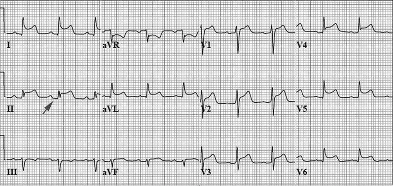
ECG is abnormal in 90% of patients with acute pericarditis and helps to distinguish it from other forms of cardiac disease, such as acute coronary syndrome. The most important ECG pattern, which reflects inflammation of the adjacent myocardium, consists of diffuse ST elevations in most ECG leads. In addition, PR-segment depression in several leads is often evident, reflecting abnormal atrial repolarization due to atrial epicardial inflammation.
The following ECG trace shows diffuse ST-segment elevation and depression of the PR segment (arrow)
Blood studies typically reveal signs of acute inflammation, including an increased white cell count (usually a mild lymphocytosis in acute viral/ idiopathic pericarditis) and elevation of serum inflammatory markers (e.g. ESR and CRP). Some patients also demonstrate elevated serum cardiac biomarkers (e.g. cardiac troponins), suggesting inflammation of the neighboring myocardium.
Further testing often includes echocardiography and additional studies that may be useful in individual cases to identify the cause of pericarditis. These tests include purified protein derivative skin test for tuberculosis, serologic tests (antinuclear antibodies and rheumatoid factor) to screen for systemic inflammatory disease, and a careful search for malignancy.
The yield of diagnostic pericardiocentesis in uncomplicated acute pericarditis is low and should be reserved for patients with very large effusions or evidence of cardiac chamber compression.
Treatment
Idiopathic or viral pericarditis is a self-limited disease that usually resolves in 1 to 3 weeks without any interventions. Management consists of rest and pain relief by analgesic and anti-inflammatory drugs. Colchicine, a drug with anti-inflammatory properties used to treat gout, may be useful as an additional agent in acute pericarditis. It has been shown to decrease the recurrence rate after an initial episode.
Oral corticosteroids are effective for severe or recurrent pericardial pain but should not be used in uncomplicated cases because of potential side effects and because steroid use is associated with an increased rate of recurrent episodes of pericarditis.
Pericarditis related to MI is also treated in a similar fashion, with rest and aspirin. Other nonsteroidal anti-inflammatory agents are often avoided immediately following an MI because of experimental evidence linking them to delayed healing of the infarct.
Purulent pericarditis requires more aggressive treatment, including catheter drainage of the pericardium and intensive antibiotic therapy. Nevertheless, mortality remains very high, even after such interventions.
Tuberculous pericarditis requires prolonged multidrug anti-tuberculous therapy.
Pericarditis in the setting of uremia often resolves following intensive dialysis.
Neoplastic pericardial disease usually indicates widely metastatic cancer, and therapy is unfortunately only palliative.
Pericardial effusion
The normal pericardial space contains 15 to 50 mL of pericardial fluid, a plasma ultrafiltrate secreted by the mesothelial cells that line the serosal layer.
A larger volume of fluid may accumulate in association with any of the forms of acute pericarditis. In addition, non-inflammatory serous effusions (transudative fluid) may result from conditions of increased capillary permeability (e.g. hypothyroidism), increased capillary hydrostatic pressure (e.g. congestive heart failure), or decreased plasma oncotic pressure (e.g. cirrhosis or nephrotic syndrome).
Chylous effusions may occur in the presence of lymphatic obstruction of pericardial drainage, most commonly caused by neoplasms and tuberculosis.
Pathophysiology
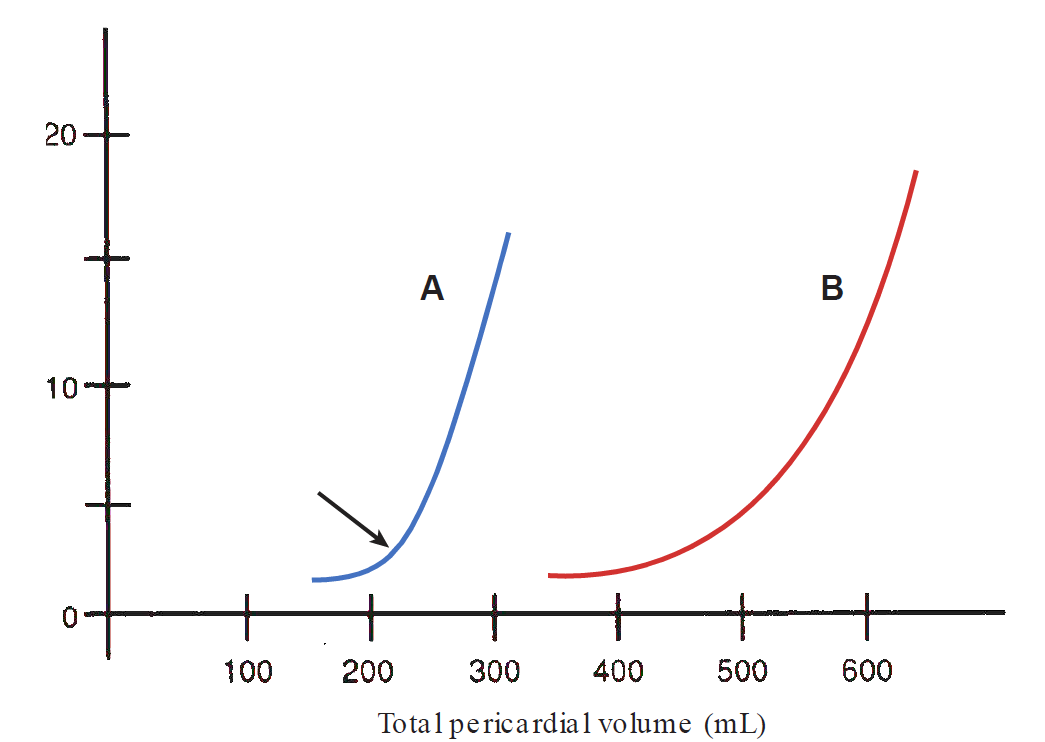
Because the pericardium is relatively stiff, the relationship between its internal volume and pressure is not linear, as shown in curve A; the initial portion of the curve is nearly flat, indicating that at the low volumes normally present within the pericardium, a small increase in volume leads to only a small rise in pressure. However, when the intrapericardial volume expands beyond a critical level (arrow), a dramatic increase in pressure is incited by the non-distensible sac. At that point, even a minor increase in volume can translate into an enormous compressive force on the heart.
Three factors determine whether a pericardial effusion remains clinically silent or whether symptoms of cardiac compression ensue:
- The volume of fluid
- The rate at which the fluid accumulates
- The compliance of the pericardium
A sudden increase in pericardial volume, as may occur in acute intrapericardial hemorrhage, results in marked elevation of pericardial pressure and the potential for severe cardiac compression. Even lesser amounts of fluid may cause significant elevation of pressure if the pericardium is pathologically noncompliant, as may occur in the presence of pericardial tumors or fibrosis. In contrast, if the pericardial effusion accumulates slowly, the pericardium gradually stretches, such that the volume–pressure relationship curve shifts toward the right (curve B in the graph above). With this adaptation, the pericardium can accommodate larger volumes without marked elevation of intrapericardial pressure or cardiac compression.
Clinical Features
A spectrum of possible symptoms is associated with pericardial effusion. For example, a patient with a large effusion may be asymptomatic, may complain of a dull constant ache in the chest, or may present with findings of cardiac tamponade. In addition, the effusion may cause symptoms of compression of adjacent structures, such as dysphagia (esophageal compression), dyspnea (lung compression), hoarseness (recurrent laryngeal nerve compression), or hiccups (phrenic nerve stimulation).
On examination, a large pericardial fluid “insulates” the heart from the chest wall, and the heart sounds may be muffled. In fact, a friction rub that had been present during the acute phase may disappear if a large effusion develops and separates the inflamed layers from one another.
Dullness to percussion of the left lung over the angle of the scapula may be present (Ewart sign) owing to compressive atelectasis by the enlarged pericardial sac.
Diagnostic studies
Chest radiography may be normal if only a small pericardial effusion is present. However, if more than approximately 250 mL has accumulated, the cardiac silhouette enlarges in a symmetric fashion.
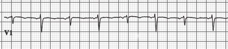
In large effusions, ECG may demonstrate reduced voltage of the complexes. In the presence of very large effusions, the height of the QRS complex may vary from beat to beat (electrical alternans) due to a constantly changing electrical axis as the heart swings from side to side within the large pericardial volume.
The following V1 ECG trace shows alternating height of the QRS complex from beat to beat
One of the most useful tests in the evaluation of effusion is echocardiography, which can identify pericardial collections as small as 20 mL. It can quantify the volume of pericardial fluid, determine whether ventricular filling is compromised, and when necessary, help direct the placement of a pericardiocentesis needle.
Treatment
If the cause of the effusion is known, therapy is directed toward the underlying disorder. If the cause is not evident, the clinical state of the patient determines whether pericardiocentesis should be undertaken.
An asymptomatic effusion, even of large volume, can be observed for long periods without specific intervention. However, if serial examination demonstrates a rise in pericardial volume or if hemodynamic compression of the cardiac chambers becomes evident, then pericardiocentesis should be performed for therapeutic drainage and analysis of the fluid.
Cardiac tamponade
At the opposite end of the spectrum from the asymptomatic pericardial effusion is cardiac tamponade. In this condition, pericardial fluid accumulates under high pressure, compresses the cardiac chambers, and severely limits filling. As a result, ventricular stroke volume and cardiac output plummets, potentially leading to hypotensive shock and death.
Any etiology of acute pericarditis can progress to cardiac tamponade, but the most common causes are neoplastic, post-viral, and uremic pericarditis. Acute hemorrhage into the pericardium is also an important cause of tamponade, which can result from blunt or penetrating chest trauma, rupture of the left ventricular free wall following MI, or as a complication of a dissecting aortic aneurysm.
Pathophysiology
As a result of the surrounding tense pericardial fluid, the heart is compressed, and the diastolic pressure within each chamber becomes elevated and equal to the pericardial pressure.
Because the compromised cardiac chambers cannot accommodate normal venous return, the systemic and pulmonary venous pressures rise. The increase of systemic venous pressure results in signs of right-sided heart failure (i.e. jugular venous distention, hepatomegaly, and peripheral edema), whereas elevated pulmonary venous pressure leads to pulmonary congestion. In addition, reduced filling of the ventricles during diastole decreases EDV, thus decreasing the stroke volume and cardiac output.
Clinical Features
Cardiac tamponade should be suspected in any patient with known pericarditis, pericardial effusion, or chest trauma who develops signs and symptoms of systemic vascular congestion and decreased cardiac output.
The key physical findings include jugular venous distention, systemic hypotension, and muffled heart sounds. These, collectively, are referred to as Beck triad. Other signs include sinus tachycardia and pulsus paradoxus.
Dyspnea and tachypnea reflect pulmonary congestion and decreased oxygen delivery to peripheral tissues.
If tamponade develops suddenly, symptoms of profound hypotension are evident, including confusion and agitation. However, if the effusion develops more gradually over weeks, then fatigue (caused by low cardiac output) and peripheral edema (owing to right-sided heart failure) may be the presenting complaints.
Pulsus paradoxus is an important sign in cardiac tamponade that can be recognized at the bedside using a standard blood pressure cuff. It refers to a decrease of systolic blood pressure by more than 10 mmHg during normal inspiration. Pulsus paradoxus is not really “paradoxical”; it is just an exaggeration of normal cardiac physiology. Normally, expansion of the thorax during inspiration causes the intrathoracic pressure to become more negative, increasing venous return and filling of the right ventricle (RV). The transient increase in RV size shifts the interventricular septum toward the left, which diminishes LV filling. As a result, in normal persons, LV stroke volume and systolic blood pressure decline slightly following inspiration.
In cardiac tamponade, this situation is exaggerated because both ventricles share a reduced, fixed volume as a result of external compression by the tense pericardial fluid. In this case, the inspiratory increase of RV volume and bulging of the interventricular septum toward the left have a proportionally greater effect on the limitation of LV filling. Thus, in tamponade, there is a more substantial reduction of LV stroke volume (and thus systolic blood pressure) following inspiration.
To measure pulsus paradoxus clinically, inflate a sphygmomanometer to a level greater than the systolic pressure. As the cuff is slowly deflated, listen for the first Korotkoff sounds, which marks the maximum systolic pressure and occurs during expiration. If the pressure is held at that level (i.e. you stop deflating the cuff) in a patient with pulsus paradoxus, the Korotkoff sounds will drift in and out, audible with expiration, and absent with inspiration (i.e. systolic pressure will fall during inspiration to a level below the cuff’s pressure and no sound will be heard). Next, slowly deflate the cuff and continue listening. When the cuff pressure falls just below the patient’s systolic pressure during inspiration, the Korotkoff sounds stop drifting in and out (i.e. they are audible during both inspiration and expiration). Pulsus paradoxus is calculated as the difference between the initial systolic pressure (when the intermittent Korotkoff sounds are first heard) and this pressure (when the sounds are first audible throughout the respiratory cycle).
Note
Pulsus paradoxus may also be manifested by other conditions in which inspiration is exaggerated, including severe asthma and chronic obstructive airway disease.
Diagnostic Studies
Echocardiography is the most useful noninvasive technique to evaluate cardiac tamponade physiology. An important indicator of high-pressure pericardial fluid is compression of the RV and right atrium during diastole. In addition, echocardiography can differentiate between cardiac tamponade and other causes of low cardiac output, such as ventricular contractile dysfunction.
The definitive diagnostic procedure for cardiac tamponade is cardiac catheterization with measurement of intracardiac and intrapericardial pressures, usually combined with therapeutic pericardiocentesis.
Treatment
Removal of the high-pressure pericardial fluid is the only intervention that reverses the physiology of this condition. Pericardiocentesis is best performed in the cardiac catheterization laboratory, where the hemodynamic effect of fluid removal can be assessed.
The patient is positioned at a 45-degree angle to promote pooling of the effusion, and a needle is inserted into the pericardial space through the skin, usually just below the xiphoid process (which is the safest location to avoid piercing a coronary artery). A catheter is then threaded into the pericardial space and connected to a transducer for pressure measurement. Another catheter is threaded through a systemic vein into the right side of the heart, and simultaneous recordings of intracardiac and intrapericardial pressures are compared.
In tamponade, the pericardial pressure is elevated and is equal to the diastolic pressures within all of the cardiac chambers, reflecting the compressive force of the surrounding effusion (this is known as equalization of cardiac pressures).
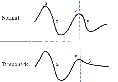
In addition, the right atrial pressure tracing, which is equivalent to the jugular venous pulsation observed on physical examination, displays a characteristic abnormality. In tamponade, the pericardial fluid compresses the RV, and so the right atrium cannot empty quickly, and the y descent in the jugular venous curve is blunted.
Following successful pericardiocentesis, the pericardial pressure falls to normal and is no longer equal to the diastolic pressures in the heart, which also decline to their appropriate levels. After initial aspiration of fluid, the pericardial catheter may be left for 1 to 2 days to allow more complete drainage. When pericardial fluid is obtained for diagnostic purposes, it should be stained and cultured for bacteria, fungi, and acid-fast bacilli (tuberculosis), and cytologic examination should be performed to evaluate for malignancy.
Other common measurements of pericardial fluid include white cell count (elevated in bacterial infections and other inflammatory conditions) and protein and lactate dehydrogenase levels. If the concentration ratio of pericardial protein to serum protein is greater than 0.5, or that of pericardial LDH to serum LDH is greater than 0.6, then the fluid is consistent with an exudate; otherwise, it is more likely a transudate.
When tuberculosis is suspected, it is also useful to measure the level of adenosine deaminase in the pericardial fluid. Studies have indicated that an elevated level is highly sensitive and specific for tuberculosis.
If cardiac tamponade recurs following pericardiocentesis, the procedure can be repeated. In some cases, a more definitive surgical undertaking (removal of part or all of the pericardium) is required to prevent reaccumulation of the effusion.
Constrictive pericarditis
The other major potential complication of pericardial diseases is constrictive pericarditis. This is a condition not frequently encountered but is important to understand, because it can masquerade as other more common disorders. In addition, it may cause profound symptoms yet is often fully correctable if recognized.
In the early part of the 20th century, tuberculosis was the major cause of constrictive pericarditis but that is much less common today. The most frequent cause now is “idiopathic” (i.e. months to years following presumed idiopathic or viral acute pericarditis). However, any etiology of pericarditis can lead to this complication.
Following an episode of acute pericarditis, any pericardial effusion that has accumulated usually undergoes gradual resorption. However, in patients who later develop constrictive pericarditis, the fluid undergoes organization, with subsequent fusion of the pericardial layers, followed by fibrous scar formation. In some patients, calcification of the adherent layers ensues, further stiffening the pericardium.
The pathophysiologic abnormalities in constrictive pericarditis occur during diastole, and systolic contraction is usually normal. In this condition, a rigid, scarred pericardium encircles the heart and inhibits normal filling. As further filling is suddenly arrested and venous return to the right heart ceases, systemic venous pressure rises, and signs of right-sided heart failure ensue. In addition, the impaired filling of the left ventricle causes a reduction in stroke volume and cardiac output, which leads to hypotension.
Clinical Features
The symptoms and signs of constrictive pericarditis usually develop over months to years. They result from reduced cardiac output (fatigue, hypotension, and reflex tachycardia) and elevated systemic venous pressures (jugular venous distention, hepatomegaly, and peripheral edema).
Because the most impressive physical findings are often the insidious development of hepatomegaly and ascites, patients may be mistakenly suspected of having hepatic cirrhosis or an intra-abdominal tumor. However, careful inspection of the elevated jugular veins can point to the correct diagnosis of constrictive pericarditis.
On cardiac examination, an early diastolic “knock” may follow S2 in patients with severe calcific constriction. It represents the sudden cessation of ventricular diastolic filling imposed by the rigid pericardial sac.
In contrast to cardiac tamponade, pericardial constriction results in pulsus paradoxus less frequently. However, in constrictive pericarditis, the negative intrathoracic pressure generated by inspiration is not easily transmitted to the RV as the non-compliant pericardium prevents the RV from expanding outward to hold the extra blood, and so inspiratory increase in RV filling is more limited. Rather, when a patient with pericardial constriction inhales, the negative intrathoracic pressure draws blood toward the thorax, accumulating in intrathoracic systemic veins, causing the jugular veins to become more distended during inspiration (known as Kussmaul sign). This is the opposite of normal physiology, in which inspiration results in a decline in jugular venous pressure as venous return is drawn into the heart.

Diagnostic Studies
Chest radiography in constrictive pericarditis shows a normal or mildly enlarged cardiac silhouette. Calcification of the pericardium can be detected in some patients with severe chronic constriction. ECG generally shows non- specific ST and T-wave abnormalities.
Echocardiographic evidence of constriction is subtle. The pericardium, if well imaged, is thickened. The ventricular cavities are small and contract vigorously, but ventricular filling terminates abruptly in early diastole, as the chambers reach the limit imposed by the surrounding rigid shell. Aberrant diastolic motion of the interventricular septum, and alterations of LV inflow velocities during respiration assessed by Doppler, also reflect the abnormal pattern of diastolic filling.
CT or MRI is superior to echocardiography in the assessment of pericardial thickness. The presence of normal pericardial thickness ( mm) by these modalities makes constrictive pericarditis a much less likely diagnosis.
The diagnosis of constrictive pericarditis can be confirmed by cardiac catheterization, which reveals four key features:
-
Elevation and equalization of the diastolic pressures in each of the cardiac chambers.
-
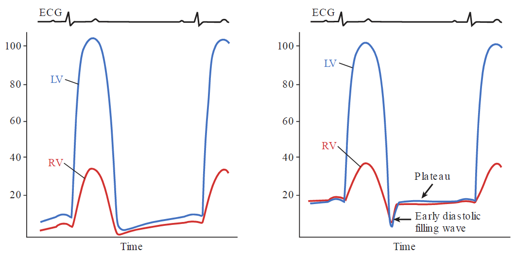
An early diastolic “dip and plateau” configuration in the RV and LV tracings. This pattern reflects blood flow into the ventricles at the very onset of diastole followed by sudden cessation of filling as further expansion of the ventricles is arrested.
-
A prominent y descent in the jugular venous pressure tracing. After the tricuspid valve opens, the right atrium quickly empties into the RV (and its pressure rapidly falls) during the very brief period before filling is arrested. This is in contrast to cardiac tamponade, in which the external compressive force prevents rapid ventricular filling, even in early diastole, such that the y descent is blunted.
-
During the respiratory cycle, there is discordance in the RV and LV systolic pressures. This is explained as follows: normally, the negative intrathoracic pressure in inspiration causes the systolic pressure of both ventricles to decline slightly. In contrast, in constrictive pericarditis, the heart is isolated from the thorax and the negative intrathoracic pressure in inspiration, which decreases the pressure in the pulmonary veins but not in the cardiac chambers. This causes a decline in the pressure gradient driving blood back to the left side of the heart from the pulmonary veins, such that left ventricle filling is diminished. Simultaneously, because the two ventricles share a fixed space limited by the rigid pericardium, the reduced LV volume allows the interventricular septum to shift toward the left, enlarging the RV (this reciprocal behavior is termed ventricular interdependence). The subsequent increase in RV filling increases its systolic pressure during inspiration. During expiration, the situation is reversed, with the RV systolic pressure declining and that of the LV increasing.
The following graphs show ventricular pressures, the first one is in a normal heart and the second is in constrictive pericarditis. In the latter situation, early diastolic ventricular filling abruptly stops as the volume in each ventricle quickly reaches the limit imposed by the pericardium. Throughout most of diastole, the LV and RV pressures are abnormally elevated and equal.
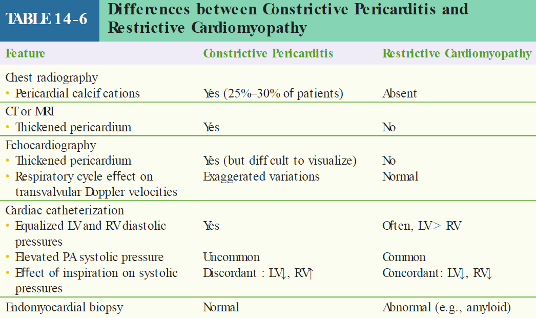
The clinical and hemodynamic findings of constrictive pericarditis are often similar to those of restrictive cardiomyopathy, another uncommon condition. Distinguishing between these is important because pericardial constriction is often correctable, whereas most cases of restrictive cardiomyopathy have very limited effective treatments. An endomyocardial biopsy is sometimes necessary to distinguish between these (biopsy is normal in constriction but usually abnormal in restrictive cardiomyopathy).
Treatment
The only effective treatment of severe constrictive pericarditis is surgical removal of the pericardium. Symptoms and signs of constriction may not resolve immediately after surgery because of the associated stiffness of the neighboring outer walls of the heart, but subsequent clinical improvement is the rule in patients with otherwise intact cardiac function. The degree of improvement depends on the underlying etiology, with the most favorable outcomes in patients with an idiopathic/post–viral pericarditis origin, and the least favorable benefit when prior radiation therapy is the cause.
See also
References
Additional Reading
- ESC Committee for Practice Guidelines. Guidelines on the diagnosis and management of pericardial diseases, executive summary. Eur Heart J. 2004;25:587–610.
- Herzog E, ed. Management of Pericardial Disease. London, UK: Springer-Verlag; 2014.
- Imazio M, Brucato A, Cemin R, et al. A randomized trial of colchicine for acute pericarditis. N Engl J Med. 2013;369:1522–1528.
- Khandaker MH, Espinosa RE, Nishimura RA, et al. Pericardial disease: diagnosis and management. Mayo Clin Proc. 2010;85:572–593.
- Klein AL, Abbara S, Agler DA, et al. American Society of Echocardiography clinical recommendations for multimodality cardiovascular imaging of patients with pericardial disease. J Am Soc Echocardiogr. 2013;26:965–1012.
- Lilly LS. Treatment of acute and recurrent idiopathic pericarditis. Circulation. 2013;127:1723–1726.
- Shabetai R. The Pericardium. Boston, MA: Kluwer Academic; 2003