Part 7: Valvular Heart Disease
Fri Jul 24 2026
By B. Hassan

Acute rheumatic fever
Acute rheumatic fever (ARF) is an inflammatory condition that affects the heart, skin, and connective tissues. Its incidence has decreased dramatically in industrialized societies, where it is now rare. However, it remains a major burden in developing countries.
ARF arises as a complication of pharyngitis caused by group A beta-hemolytic streptococci and mainly affects children and young adults. During prior epidemics, approximately 3% of patients with acute streptococcal pharyngitis developed ARF 2-3 weeks after the initial throat infection.
Common presenting symptoms are chills, fever, fatigue, and migratory arthritis. The clinical manifestations that establish the diagnosis are known as Jones criteria.
Involvement of the heart results from autoimmune cross-reactivity between the bacterial and cardiac antigens. Pathologically, carditis affects all layers of the heart (pericardium, myocardium, and endocardium). During the acute episode, carditis may cause tachycardia, impaired ventricular contractility, a pericardial friction rub, and transient murmurs that reflect turbulent flow across inflamed valve leaflets.
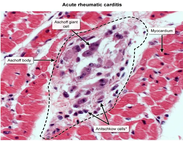
Histopathologic examination may demonstrate Aschoff bodies, areas of local fibrinoid necrosis surrounded by inflammatory cells that later resolve to form fibrous scar tissue.
The most important sequela of ARF is chronic rheumatic heart disease (RHD) characterized by permanent deformity and impairment of one or more cardiac valves. Symptoms of valvular dysfunction, however, do not manifest until 10 to 30 years after ARF has subsided. This latency period may be shorter with more aggressive disease. RHD affects the mitral valve in almost all cases, the aortic valve in 20% to 30%, and rarely the tricuspid valve. Stenosis and/or regurgitation of each valve can result.
Treatment of acute infection includes high-dose aspirin to reduce inflammation and penicillin to eliminate residual streptococcal infection. Management of RHD, on the other hand, includes prophylaxis against recurrent streptococcal infection and treatment of the chronic valve lesions. Recurrences of ARF can precipitate further cardiac damage, so individuals with ARF should receive low-dose penicillin prophylaxis at least until early adulthood, by which time exposure and susceptibility to streptococcal infections have diminished.
Jones criteria
Major criteria:
-
Carditis (inflammation of all 3 heart layers)
-
Migratory arthritis (mainly affecting large joints)
-
Sydenham chorea (involuntary movements affecting proximal muscles more than distal muscles and associated with hypotonia and hyporeflexia)
-
Erythema marginatum (painless red skin nodules with serpiginous margins that gradually enlarge while the center becomes pale)
-
Subcutaneous nodules
Minor criteria:
- Arthralgias
- Fever
- Elevated acute-phase reactants (ESR, CRP)
- Prolonged PR interval on ECG
Evidence of group A streptococcal infection can be obtained using antistreptolysin O (ASO) antibodies or through a positive throat culture or rapid antigen test.
Diagnosis requires evidence of streptococcal infection and either 2 major criteria or 1 major plus 2 minor criteria.
The following image shows erythema marginatum:

Endocarditis
Infection of the endocardial surface, including the cardiac valves, can lead to extensive tissue damage, which may be fatal. Infective endocarditis (IE) carries an overall 6-month mortality rate of 20% to 25%, even with appropriate therapy, and a 100% mortality rate if it is not recognized and treated correctly.
There are 3 ways to classify IE:
- By clinical course
- By host substrate
- By the specific infecting microorganism
In the first classification scheme, IE is termed acute bacterial endocarditis (ABE) when the infection presents as an acute fulminant infection, and the causative organism is a highly virulent and invasive organism such as Staphylococcus aureus. When IE presents with a more insidious clinical course, it is termed subacute bacterial endocarditis (SBE) and the causative organism is typically less virulent like Streptococci viridans.
Note
Because of the virulence of the responsible microorganism, ABE may occur on previously healthy heart valves. On the other hand, SBE most frequently occurs in individuals with underlying valvular damage.
The second means of classification is according to the host substrate. This includes native valve endocarditis, prosthetic valve endocarditis, or endocarditis in the setting of IV drug abuse. Of these, native valve endocarditis accounts for 60% to 80% of patients. Different microorganisms and clinical courses are associated with each of these categories. For example, the skin contaminant Staphylococcus epidermidis is a common cause of prosthetic valve endocarditis, but rarely is the cause of native valve endocarditis. IV drug users have a propensity for S. aureus endocarditis of the right-sided heart valves.
The third classification is according to the specific infectious agent. The most common responsible organisms are gram-positive cocci. Certain bacterial strains that cause endocarditis are associated with particular anatomic sources. For example, viridans group streptococci usually originate from oropharyngeal tissue, while Streptococcus bovis (more recently termed S. gallolyticus) commonly arises from the gastrointestinal tract and should prompt investigation for colonic polyps or adenocarcinoma.
Pathogenesis
The course of endocarditis usually follows specific events, requiring several conditions to occur. First, the endothelial surface has to be injured, then a thrombus has to form on the site of endothelial injury. This provides a favorable environment for infection. Finally, bacteria have to enter into the circulation and colonize the injured endocardial surface.
The most common cause of endothelial injury is turbulent blood flow resulting from preexisting cardiac or intravascular abnormalities, including acquired valvular heart lesions, congenital heart diseases, and hypertrophic cardiomyopathy. Endothelial injury may also be incited by foreign material within the circulation, such as indwelling venous catheters, IV drug abuse, prosthetic heart valves, and other implanted cardiac devices.
Once an endocardial surface is injured, platelets adhere to the exposed subendocardial connective tissue and initiate the formation of a sterile thrombus (termed a vegetation) through fibrin deposition. This process is referred to as nonbacterial thrombotic endocarditis (NBTE), which makes the endocardium more hospitable to microbes by providing a surface for attachment and also protects adherent organisms from immune defenses by inhibiting chemotaxis.
Note
The normal endocardial surface is usually very smooth such that it prevents any bacterial attachment. However, NBTE impairs that smoothness and allows for bacterial attachment.
Three factors determine the ability of a microorganism to induce IE: access to the bloodstream, survival in the circulation, and adherence to the endocardium. Bacteria can be introduced into the bloodstream whenever a mucosal or skin surface harboring an organism is injured, such as from the mouth during dental procedures, from the skin during IV drug use, or from an indwelling venous catheter. However, while transient bacteremia is relatively common, not all bacteria can survive in the circulation or adhere to a fibrin thrombus. For example, gram-positive organisms account for the majority of cases of endocarditis because of their resistance to destruction in the circulation by the complement system. Moreover, many streptococcal species tend to produce dextran, a bacterial cell wall component that adheres to thrombi, which correlates with their ability to cause endocarditis.
Once organisms adhere to the injured surface and are protected from phagocytic activity by the overlying fibrin, they multiply rapidly, enlarging the infected vegetation. As the vegetation serves as a source of continuous bacteremia, many complications can occur, including septic emboli, cardiac injury, and immune injury mediated by antigen–antibody deposition. For example, local extension of the infection within the heart can result in progressive valve damage, abscess formation, or erosion into the cardiac conduction system. Additionally, immune complex deposition can result in glomerulonephritis, arthritis, or vasculitis.
The epidemiology of IE has evolved in recent decades as bacteria resistant to antibiotics have become ubiquitous in the hospital setting and have spread into the community. Antibiotic resistant strains such as methicillin-resistant S. aureus and vancomycin-resistant enterococci have become more common and are associated with increased mortality rates from IE.
Clinical Manifestations
A patient with acute IE is likely to report an explosive and rapidly progressive illness with high fever and shaking chills. In contrast, subacute IE presents less dramatically with low-grade fever often accompanied by non-specific constitutional symptoms such as fatigue, anorexia, weakness, myalgia, and night sweats. Other symptoms include splenomegaly, elevated white blood cell count with a left shift (increased proportion of neutrophils and immature granulocytes), elevated inflammatory markers like ESR and CRP, and in approximately 50% of cases, an elevated serum rheumatoid factor.
These symptoms could easily be mistaken for influenza or an upper respiratory tract infection. Thus, the diagnosis of subacute IE requires a high index of suspicion. A history of a valve lesion or other condition known to predispose to endocarditis is helpful. A thorough history should also inquire about IV drug use, recent dental procedures, or other potential sources of bacteremia.
Cardiac examination may reveal a murmur representing underlying valvular pathology that predisposed the patient to IE, or a new murmur due to IE-induced damage. The development of right-sided valve lesions (e.g. tricuspid regurgitation), although rare in normal hosts, is particularly common in endocarditis associated with IV drug abuse. During the course of endocarditis, severe valvular destruction may result in signs of heart failure, which is the leading cause of death in patients with IE.
Other physical findings that may appear in IE are those associated with septic embolism or immune complex deposition. Brain emboli occur in up to 40% of patients, often resulting in new neurologic findings on physical examination. Renal injury may also present with flank pain, hematuria, or acute renal failure. Lung infarction (septic pulmonary embolism) or infection (pneumonia) is particularly common in endocarditis that involves right-sided valves. Embolism of the vasa vasorum of arteries can cause localized aneurysm formation (termed a mycotic aneurysm), and is particularly dangerous in cerebral vessels, because rupture there can result in fatal intracranial hemorrhage.
Skin findings resulting from septic embolism or immune-complex vasculitis are often collectively referred to as peripheral stigmata of endocarditis. These include petechiae (tiny, circular, red-brown discolorations on mucosal surfaces or skin) and splinter hemorrhages (longitudinal hemorrhages found under the nails) due to micro-emboli.
Other peripheral stigmata are now rarely encountered but include Janeway lesions, which are painless, flat, irregular discolorations on the palms and soles; Osler nodules, which are tender erythematous nodules found primarily in the pulp space of the fingers and toes; Roth spots, representing micro-emboli to the retina, and appear as white dots surrounded by hemorrhage on fundal examination.
ECG may help identify extension into the cardiac conduction system, manifesting as heart block or new arrhythmias. Echocardiography is used to visualize vegetations, valvular dysfunction, and abscess formation. Transthoracic echocardiography is useful in detecting large vegetations and has the advantage of being non-invasive. However, while its specificity for vegetations is high, its sensitivity is less than 60%. Transesophageal echocardiography, on the other hand, is much more sensitive (> 90%) for detecting vegetations and myocardial abscesses and can be particularly useful for the evaluation of infection involving prosthetic valves.
Central to the diagnosis and treatment is a blood culture to identify the causative organism, allowing treatment to be tailored to the causative organism. A specific etiologic agent is identified approximately 90% of the time. However, blood cultures may return negative if antibiotics have already been administered or if the organism has unusual growth requirements. Even after a careful history, examination, and evaluation of laboratory data, the diagnosis of IE can be elusive. Therefore, attempts have been made to standardize the diagnosis, resulting in the widely used Duke criteria, where diagnosis of endocarditis requires the presence of two major criteria, one major and three minor criteria, or five minor criteria.
Treatment
Treatment of endocarditis involves 4-6 weeks of high-dose antibiotic therapy. Although empiric broad-spectrum antibiotics may be used before blood cultures are obtained for severely ill patients, switch to specific directed therapy should occur once the causative microorganism has been identified.
Surgical intervention with valve replacement is indicated for patients with persistent bacteremia despite appropriate antibiotic therapy, those with severe valvular dysfunction leading to heart failure, or for individuals with myocardial abscesses or recurrent endocarditis-related thrombo-emboli.
Prevention
Prevention of endocarditis requires administering antibiotics to susceptible individuals before invasive procedures that are likely to result in bacteremia.
Cardiac conditions for which antibiotic prophylaxis is reasonable:
-
Patients with a prosthetic heart valve or prior valve repair with prosthetic material
-
Prior history of endocarditis
-
Certain congenital heart diseases (CHD) like unrepaired cyanotic CHD, repaired CHD with prosthetic material, during the first 6 months after the procedure
-
Cardiac transplant recipients who develop cardiac valve abnormalities
Procedures that warrant antibiotic therapy for conditions listed above:
-
Dental procedures that involve manipulation of gingival tissue, manipulation of periapical region of the teeth, or perforation of the oral mucosa
-
Upper respiratory tract procedures, only if involves incision or biopsy of mucosa (e.g. tonsillectomy, bronchoscopy with biopsy)
-
Genitourinary or gastrointestinal procedures, only if infections of those systems are present
-
Procedures on infected skin or musculoskeletal tissue
Mitral Stenosis
The most common underlying cause of mitral stenosis (MS) is prior rheumatic fever. Approximately 50% to 70% of patients with symptomatic MS have a history of acute rheumatic fever, on average, 20 years before presentation. Other rare etiologies of MS include calcification of the valve, infective endocarditis with large vegetations that obstruct the valve orifice, and rare congenital stenosis of the valve.
Pathologic features of MS include fibrous thickening and calcification of the valve leaflets, fusion of the commissures, and thickening and shortening of the chordae tendineae.
Pathophysiology
In MS, there is obstruction to blood flow across the valve such that emptying of the left atrium is impeded and there is an abnormal pressure gradient between the left atrium and the left ventricle. As a result, the left atrial pressure increases.
The high left atrial pressure is transmitted retrograde to the pulmonary circulation, resulting in pulmonary hypertension, which may cause transudation of plasma fluids into the lung interstitium, causing dyspnea and other symptoms of heart failure. In severe cases, significant elevation of pulmonary venous pressure leads to a fistula between the pulmonary and bronchial veins. Subsequently, an engorged bronchial vein may rupture into a bronchus, causing hemoptysis (coughing up blood).
The elevation of left atrial pressure can result in two distinct types of pulmonary hypertension: passive and reactive. Most patients with MS exhibit passive pulmonary hypertension, related to the backward transmission of the elevated LA pressure into the pulmonary vasculature as described above. This represents an “obligatory” increase in pulmonary artery pressure that preserves forward flow in the setting of increased left atrial and pulmonary venous pressures.
Approximately 40% of patients with MS demonstrate reactive pulmonary hypertension with medial hypertrophy and intimal fibrosis of the pulmonary arterioles. This initially serves a beneficial role because the increased arteriolar resistance impedes blood flow into the engorged pulmonary capillary bed and thus reduces capillary hydrostatic pressure. However, this benefit is at the cost of elevation of the right-sided heart pressures, as the right ventricle pumps against the increased resistance. Chronic elevation of right ventricular pressure leads to hypertrophy and dilatation of that chamber and ultimately to right-sided heart failure.
Chronic pressure overload of the LA in MS leads to left atrial enlargement and dilation, stretching the fibers of the electro-conductive system located in the left atrium, resulting in atrial fibrillation. The relative stagnation of blood in the dilated LA, especially when combined with the development of atrial fibrillation, predisposes to intra-atrial thrombus formation, which may serve as an embolism to other organs, leading to severe complications like a stroke. Thus, MS patients who develop atrial fibrillation require chronic anticoagulation therapy.
Left ventricular pressures are usually normal, but impaired ventricular filling through the stenotic valve may reduce LV stroke volume and cardiac output.
Presentation
The 10-year survival exceeds 80% in asymptomatic or minimally symptomatic patients. However, the 10-year survival of untreated patients after onset of symptoms is only 50-60%. Longevity is much more limited for patients with advanced symptoms and is abysmal for those who develop significant pulmonary hypertension, with a mean survival of less than 3 years.
The clinical presentation of MS depends on the degree of reduction of the valve area. The earliest manifestations are those of dyspnea and reduced exercise capacity. In mild MS, dyspnea may be absent at rest; however, it develops on exertion as the exercise-induced increased heart rate decreases diastolic filling time. Other conditions and activities that increase heart rate and precipitate or exacerbate symptoms of MS include fever, anemia, hyperthyroidism, pregnancy, rapid arrhythmias, emotional stress, and sexual intercourse.
With more severe MS, dyspnea occurs even at rest. Increasing fatigue and more severe signs of pulmonary congestion, such as orthopnea and paroxysmal nocturnal dyspnea occur. With advanced MS and pulmonary hypertension, signs of right-sided heart failure may occur, including jugular venous distention, hepatomegaly, ascites, and peripheral edema. Compression of the recurrent laryngeal nerve by an enlarged pulmonary artery or left atrium may cause hoarseness (known as Ortner syndrome).
Less often, the diagnosis of MS is made by one of its complications: atrial fibrillation, thromboembolism, infective endocarditis, or hemoptysis.
Examination
On examination, there are several typical findings in MS. Auscultation shows a loud S1 in the early stages of the disease, resulting from the high LA pressures, which keeps the valve leaflets separated throughout diastole; at the onset of systole, ventricular contraction abruptly slams the leaflets from a relatively wide position, causing the closure sound to be more prominent. In late stages of the disease, the intensity of S1 may normalize or become reduced as the valve leaflets thicken, calcify, and become less mobile.
A main feature of auscultation in MS is an opening snap (OS) following S2. The OS is thought to result from the sudden tensing of the chordae tendineae and stenotic leaflets on opening of the abnormal valve. The interval between S2 and the OS relates inversely to the severity of MS; the more severe the MS, the higher the LA pressure and the earlier the valve is forced open in diastole. The OS is followed by a low-frequency decrescendo murmur (termed diastolic rumble) caused by turbulent flow across the stenotic valve during diastole. The duration, but not the intensity, of the diastolic murmur relates to the severity of MS. The more severe the stenosis, the longer it takes for the LA to empty and for the gradient between the LA and LV to dissipate.
At the end of diastole, contraction of the LA causes the pressure gradient between the LA and LV to rise again; therefore, the murmur briefly becomes louder at that time (termed presystolic accentuation). This final accentuation of the murmur does not occur in atrial fibrillation because there is no effective atrial contraction in that situation.
Murmurs caused by other valve lesions are often found concurrently in patients with MS. For example, mitral regurgitation frequently coexists with MS. Additionally, right-sided heart failure caused by severe MS may induce tricuspid regurgitation as a result of right ventricular enlargement. Moreover, aortic regurgitation (because of rheumatic involvement of the aortic leaflets) or pulmonic regurgitation (because of MS-induced pulmonary hypertension) may also be present.
An ECG in MS shows left atrial enlargement and, if pulmonary hypertension occurs, right ventricular hypertrophy. Atrial fibrillation may also be present.
A chest X-ray may reveal left atrial enlargement, interstitial edema, and Kerley B lines resulting from pulmonary edema. With the development of pulmonary hypertension, right ventricular enlargement and prominence of the pulmonary arteries appear.
Echocardiography is of major diagnostic value in MS. Structural findings include thickened mitral leaflets with abnormal fusion of their commissures and restricted separation during diastole. The degree of left atrial enlargement can be quantified, and if present, intra-atrial thrombus may be visualized. The mitral valve area can be measured directly on cross-sectional views or calculated from Doppler velocity measurements (a technique known as “diastolic pressure half-time”).
Patients can be stratified into stages of severity based on the mitral valve area. A normal mitral valve orifice measures between 4 and 6 . Current guidelines define clinically important “severe” MS as a valve area , a state that is typically accompanied by LA enlargement and elevated pulmonary artery systolic pressure. A valve area is considered “very severe” MS. If the findings determined by echocardiography seem milder than the patient’s history and examination suggest, an exercise test with accompanying Doppler assessment, or cardiac catheterization may be warranted.
Treatment
Salt restriction and diuretic therapy may improve symptoms due to vascular congestion. Heart rate slowing agents, such as β-blockers or non-dihydropyridine calcium channel blockers (e.g. diltiazem or verapamil) increase diastolic LV filling time and therefore ease symptoms that occur during exercise.
Anticoagulant therapy to prevent thromboembolism is recommended for MS patients with atrial fibrillation, or an identified atrial thrombus, or prior embolic events.
Percutaneous or surgical valve interventions are the only treatments that alter the prognosis of MS and are indicated in patients with severe, symptomatic MS. Percutaneous balloon mitral valvuloplasty is the treatment of choice in appropriately selected patients (those without advanced anatomic valve deformity, mitral regurgitation, or left atrial thrombus). During this procedure, a balloon catheter is advanced from the femoral vein into the right atrium, then across the atrial septum by intentionally puncturing the interatrial septum, and then through the narrowed mitral valve orifice. The balloon is then inflated, opening the valve commissures.
In young adults with the suitable anatomy for the procedure, the event-free survival rate approaches 80% to 90% over 3 to 7 years of follow-up. Approximately 5% of patients undergoing balloon mitral valvuloplasty are left with a residual atrial septal defect due to the transseptal puncture. Less common complications include liberation of emboli at the time of valvuloplasty, cardiac perforation by the catheter, or the unintentional creation of substantial mitral regurgitation.
Open mitral valve commissurotomy is an operation in which the stenotic commissures are separated under open heart surgery. It may be undertaken in patients for whom percutaneous balloon valvuloplasty is not feasible. It is effective in relieving obstruction, and restenosis occurs in fewer than 20% of patients over 10 to 20 years of follow-up. Perioperative mortality rates are low (2%).
Mitral valve replacement is considered in patients who are not appropriate candidates for balloon valvuloplasty or open commissurotomy.
Mitral Regurgitation
The mitral valve is a complex structure composed of an annulus, two leaflets, chordae tendineae, and papillary muscles, supported by the adjacent myocardium to which the annulus and papillary muscles are attached. Disruption to any of these components can result in abnormal closure of the valve during systole, causing mitral regurgitation (MR).
MR is categorized as primary if it occurs due to a structural defect of one or more valve components, or secondary if the valve is structurally normal, but regurgitation instead results from left ventricular enlargement. In the latter case, dilatation of the mitral annulus by the enlarged LV, and/or spatial separation of the papillary muscles causes abnormal closure of the valve, leading to the development of MR.
MR can also present as an “acute” or “chronic” condition, with different pathophysiologic consequences. Most cases of acute MR are primary in nature and result from sudden damage to components of the valve apparatus. For example, rupture of an infarcted papillary muscle can occur within days of a STEMI, often resulting in severe MR. Acute MR due to sudden rupture of chordae tendineae can result from infective endocarditis, chest trauma, or from degeneration of the chordae owing to connective tissue disorders such as Marfan syndrome.
Chronic MR has multiple primary causes, including myxomatous degeneration of the valve, in which “floppy” leaflets allow regurgitation to occur by bowing excessively into the LA during systole (termed mitral valve prolapse). Other causes of chronic primary MR include rheumatic deformity of the valve, congenital valve defects, and extensive calcification of the mitral annulus.
Pathophysiology
In MR, a portion of the left ventricular stroke volume is ejected backward into the LA during systole. As a result, the forward cardiac output (into the aorta) is less than the LV’s total output. Therefore, the direct consequences of MR include elevation of left atrial volume and pressure; a reduction of forward cardiac output; and a volume-related stress on the LV because the regurgitant volume returns to the LV in diastole along with the normal pulmonary venous return.
To meet normal circulatory needs and to eject the additional volume, LV stroke volume must rise. This increase is accomplished by the Frank–Starling mechanism, whereby the elevated LV diastolic volume increases myofiber stretch, increasing the stroke volume.
The hemodynamic consequences of MR vary depending on the degree of regurgitation and how long it has been present.
The regurgitant fraction in MR is defined as:
This ratio rises whenever the resistance in the aorta is increased, as blood follows the path of least resistance. For example, high systemic blood pressure or the presence of aortic stenosis will increase the regurgitant fraction.
The extent to which left atrial pressure rises in response to the regurgitant volume is determined by the left atrial compliance. Compliance is a measure of the chamber’s pressure–volume relationship, reflecting the ease or difficulty with which the chamber can be filled.
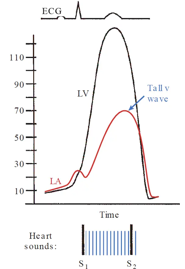
In acute MR, left atrial compliance undergoes little change. Because the LA is a relatively stiff chamber, its pressure increases substantially when it is suddenly exposed to regurgitant blood. This elevated pressure is transmitted backward to the pulmonary circulation and can result in rapid pulmonary congestion and edema. In this case, the LA pressure or the pulmonary capillary wedge pressure demonstrates a prominent v wave (often referred to as a “cv” wave when it merges with the preceding c wave), reflecting the increased LA filling during systole. Additionally, as in MS, pulmonary artery and right-heart pressures passively rise.
In acute MR, the LV accommodates the increased end diastolic volume (volume load returning from the LA) according to the Frank–Starling relationship. The result is a compensatory increase in the LV stroke volume such that the left ventricular end systolic volume remains normal. Moreover, left ventricular emptying is facilitated by the reduced total impedance to LV contraction (i.e. the afterload decreases), since a portion of the LV output is directed into the low-impedance LA, rather than into the higher-pressure aorta.
In contrast to the acute situation, the gradual course of chronic MR permits the LA to undergo compensatory changes. In particular, the LA compliance increases such that the chamber is able to accommodate a larger volume without a substantial increase in pressure. However, this adaptation occurs at the cost of reduced forward cardiac output, because the compliant LA becomes a preferred low-pressure “sink” for the left ventricle compared with the aorta. Consequently, as progressively larger fractions of blood regurgitate into the LA, chronic MR causes symptoms of low cardiac output (e.g. weakness and fatigue). In addition, chronic left atrial dilatation predisposes to the development of atrial fibrillation.
In chronic MR, the LV also undergoes gradual dilation and eccentric hypertrophy in response to the increased end diastolic volume, increasing the ventricular compliance to accommodate the increased filling volume. Forward output is preserved to near-normal levels for many years by maintaining a higher stroke volume via the Frank–Starling mechanism. Over years, however, chronic eccentric hypertrophy results in overstretching, causing deterioration of systolic ventricular function, a decline in forward output, and symptoms of heart failure.
Examination
Auscultation of a patient with chronic MR typically reveals an apical holosystolic (pansystolic) murmur that often radiates to the axilla. This description has several exceptions. In patients with severe acute MR, the character of the systolic murmur is often different, occurring in early to mid systole with a decrescendo quality. In isolated posterior mitral leaflet prolapse, the regurgitant jet is directed anteriorly, and the murmur may instead radiate to the base of the heart and could be confused with the murmur of aortic stenosis (AS) in that location.
Fortunately, the distinction between the murmur of MR and that of AS can be made by simple bedside maneuvers. If the patient clenches their fists and forearms, systemic vascular resistance increases and the murmur of MR will intensify, whereas the murmur of AS will not.
Even more helpful in this distinction is the effect of the time between consecutive heart beats on the intensity of the murmur. As the length between 2 consecutive beats increases, LV filling also increases. The intensity of the murmur of AS is directly proportional to the length of the preceding cycle. This is because as LV filling increases, even small pressure gradients are amplified as more blood is ejected across the stenotic valve. In MR, however, the intensity of the murmur does not vary significantly because the change in the LV-LA pressure gradient is minimally affected by alterations in the cycle length.
In addition to the systolic murmur, a common finding in chronic MR is an S3, which reflects increased volume returning to the LV in early diastole. Additionally, in chronic MR, the cardiac apical impulse is often laterally displaced toward the axilla because of LV enlargement.
Echocardiography can often identify the cause of the MR and assess its severity. Chest radiograph may show pulmonary edema in acute MR but chronic asymptomatic MR more likely demonstrates LV and LA, without pulmonary congestion.
Left ventriculography can confirm the findings, showing a large v wave in the pulmonary capillary wedge pressure tracing (reflective of LA pressure) in acute cases. The v wave becomes less prominent, however, with progressive LA dilatation and greater compliance over time.
The following graph shows the hemodynamic profile of MR. A large systolic v wave is noted in the LA pressure tracing. A holosystolic murmur is present in chronic MR (as shown here), beginning at S1 and continuing through S2.
Treatment
Acute severe MR is a surgical emergency with a poor prognosis, even with appropriate treatment, with a 30-day mortality rate of 20% to 25%. Pharmacologic therapy is useful only to stabilize patients until surgery. For example, IV nitroprusside decreases arterial resistance, increasing forward flow and diminishing the regurgitant volume. In this way, cardiac output and pulmonary congestion may improve transiently.
The course of the disease differs depending on the cause. For example, in RHD, the course is of very slow progression with a 15-year survival rate of 70%. On the other hand, abrupt worsening of chronic MR of any cause can occur with superimposed complications such as rupture of chordae tendineae or endocarditis, and can result in an immediate life-threatening situation.
In chronic primary MR, medical treatment with vasodilators is less useful than in acute MR and has not been shown to delay the need for valve surgery in chronic MR.
Surgical intervention should be undertaken in symptomatic patients, or at the earliest sign of LV contractile dysfunction on imaging (i.e. a fall in EF to by echocardiography) even before symptoms develop. Surgical intervention consists of either mitral valve repair or replacement, depending on the underlying cause and valve anatomy.
Surgical options for chronic MR include mitral valve repair or replacement. Mitral valve repair is the preferred operative technique when feasible, as it eliminates many of the problems associated with artificial valves. It involves the reconstruction of the damaged parts of the valve. For example, a perforated leaflet may be patched with autologous pericardium transplant, or ruptured chordae may be reattached to a papillary muscle.
Operative mortality rates for patients with MR in the Society for Thoracic Surgeons database are less than 2% for mitral valve repair and 5% to 7% for mitral valve replacement. These rates are higher if concurrent coronary artery bypass grafting is performed. In general, mitral valve repair is more often appropriate for younger patients with myxomatous involvement of the mitral valve, and mitral replacement is more often undertaken in older patients with more extensive valve pathology.
In patients with chronic, severe, primary MR who are at prohibitive operative risk, a recently developed technique of transcatheter mitral valve repair can be considered. In this procedure, a catheter is advanced from the femoral vein into the right atrium, then into the left via a puncture through the interatrial septum (similar to mitral balloon valvuloplasty), and advanced into the left ventricle. A mechanical clip is then used to grasp and tether the anterior and posterior mitral leaflets and is then left in place. The procedure has been shown to be safe and effective. However, in a randomized trial of transcatheter repair versus valve surgery for patients with severe primary MR, surgery proved more effective and remains the intervention of choice in patients who are acceptable candidates for an operation.
Because chronic, secondary MR is often a result of left ventricular dysfunction, pharmacologic intervention is the treatment of choice, using a standard combination of heart failure medications, including diuretics, ACE inhibitors or angiotensin receptor blockers, β-blockers, and aldosterone antagonists. Surgical intervention is considered only when a patient with chronic, severe secondary MR has persistent symptoms despite optimal medical therapy.
Mitral Valve Prolapse
Mitral valve prolapse (MVP) is characterized by abnormal bowing of a portion of one or both mitral leaflets into the LA during ventricular systole, and is frequently accompanied by MR. Other names for this condition include floppy mitral valve, myxomatous mitral valve, and Barlow syndrome.
MVP may be inherited as an autosomal dominant disorder with variable penetrance, or it may accompany certain connective tissue diseases such as Marfan syndrome or Ehlers–Danlos syndrome. A recent echocardiographic study indicated that MVP occurs in about 2% of the population and is more common among women, especially those who are thin.
Pathologically, the valve leaflets, particularly the posterior leaflet, are enlarged, and the normal dense collagen and elastin matrix of the valvular cusps are fragmented and replaced with loose myxomatous connective tissue. Additionally, in more severe lesions, elongated or ruptured chordae, annular enlargement, or thickened leaflets may be present.
MVP is often asymptomatic but affected individuals may describe chest pain or palpitations because of associated arrhythmias. Most often it is identified on routine physical examination by the presence of a mid-systolic click and late systolic murmur heard best at the cardiac apex. The click is thought to correspond to the sudden tensing of the involved mitral leaflet or chordae tendineae as the leaflet is forced toward the LA; the murmur corresponds to regurgitant flow through the incompetent valve.
The click and murmur are characteristically altered using some bedside maneuvers. Maneuvers that increase the volume of the LV (e.g. squatting) place traction on the chordae tendineae, limiting and delaying the occurrence of prolapse and cause the click and murmur to occur later. Conversely, maneuvers that decrease the volume in the LV (e.g. sudden standing) makes the prolapse occur earlier, and the click and murmur move closer to S1.
Confirmation of the diagnosis is obtained by echocardiography. ECG and chest radiographs are usually normal unless chronic MR has resulted in left atrial and left ventricular enlargement.
The clinical course of MVP is most often benign. Treatment consists of reassurance about the good prognosis and monitoring for the development of progressive MR. Occasionally, rupture of myxomatous chordae can cause sudden severe regurgitation and pulmonary edema. Other potential complications include infective endocarditis, peripheral emboli due to microthrombus formation on the redundant valve tissue, and arrhythmias.
Aortic Stenosis
There are 3 major causes of aortic stenosis (AS):
- Degenerative calcification of a normal trileaflet aortic valve
- Calcification of a congenital bicuspid aortic valve
- Rheumatic aortic valve disease
Bicuspid aortic valves are present in 1-2% of the population and such patients typically develop signs of severe valve disease about a decade earlier than patients with the normal trileaflet type of AS. Rheumatic aortic valve disease is now uncommon in developed countries and is nearly always accompanied by rheumatic involvement of the mitral valve.
Pathology
The pathologic appearance in AS is dependent on its etiology. Degenerative, calcific AS results from endothelial dysfunction, lipid accumulation, inflammation, and alteration of signaling pathways that appears similar to atherogenesis as we talked about earlier. Over time, valvular fibroblasts differentiate into osteoblast-like cells that deposit calcium hydroxyapatite crystals on the valve, resulting in leaflet thickening and stiffening. This process is likely exacerbated by abnormal shearing forces, as occur with congenital bicuspid valves, and could explain the earlier presentation of such patients. As with atherosclerosis, risk factors for calcific degenerative AS include dyslipidemia, smoking, and hypertension.
In rheumatic AS, endocardial inflammation leads to organization and fibrosis of the valve and ultimately to fusion of the commissures and formation of calcified masses within the aortic cusps.
Pathophysiology
Since the obstruction in AS develops gradually, the LV is able to compensate by undergoing concentric hypertrophy in response to the increased pressure load. Initially, such hypertrophy serves an important role in reducing LV wall stress. Over time, however, it also reduces the compliance of the ventricle (i.e. diastolic failure).
In normal individuals, left atrial contraction contributes only a small portion of the left ventricular filling. In patients with hypertrophic cardiomyopathy and aortic stenosis, the stiffened LV becomes dependent on atrial contraction to maintain adequate ventricular filling (atrial contraction provides more than 25% of the stroke volume to the stiffened LV). This is called atrial kick, and without atrial contraction (e.g. in atrial fibrillation), decreased preload can cause marked deterioration, including severe hypotension and pulmonary edema.
Three major manifestations occur in patients with advanced AS: angina, exertional syncope, and heart failure. Each manifestation, in order, heralds an increasingly worse prognosis.
AS may result in angina because it creates an imbalance between myocardial oxygen supply and demand. Myocardial oxygen demand increases as the hypertrophied LV requires greater-than-normal perfusion. In addition, AS reduces myocardial oxygen supply because the elevated left ventricular diastolic pressure reduces the coronary perfusion pressure gradient between the aorta and the myocardium.
Exertional syncope occurs because although left ventricular hypertrophy allows the chamber to generate a high pressure and maintain cardiac output at rest, the ventricle cannot significantly increase its cardiac output during exercise because of the fixed stenotic aortic orifice. This causes decreased cerebral perfusion and, potentially, loss of consciousness on exertion.
Finally, AS can result in symptoms of heart failure. With progression of the stenosis, the LV may develop contractile dysfunction because of the high afterload, leading to increased left ventricular diastolic volume and pressure. The accompanying marked elevation of LA and pulmonary venous pressures incites pulmonary alveolar congestion and symptoms of heart failure.
Examination
The key features of advanced AS include a coarse crescendo–decrescendo systolic ejection murmur with a weakened (parvus) and delayed (tardus) upstroke of the carotid artery owing to the obstructed LV outflow.
Other common findings on cardiac examination include the presence of an S4 because of atrial contraction into the stiff LV and reduced intensity, or complete absence, of the A2 component of S2.
A normal aortic valve has a cross-sectional area of 3-4 and a mean systolic pressure gradient between the LV and aorta of less than 5 mmHg. As the valve area decreases, the pressure gradient rises. When the valve area declines to , or the mean pressure gradient increases to greater than 40 mmHg, a patient is considered to have severe aortic stenosis and symptoms typically appear.
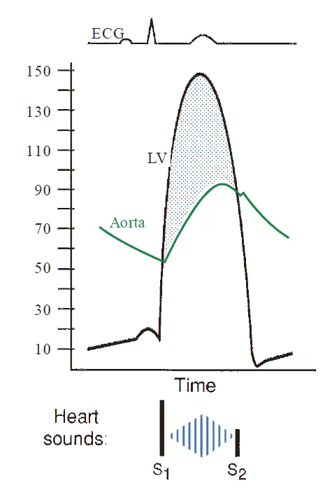
The following graph shows the hemodynamic profile of AS. A systolic pressure gradient (shaded area) is present between the LV and aorta. S2 is diminished in intensity, and there is a crescendo–decrescendo systolic murmur that does not extend beyond S2.
An ECG may show left ventricular hypertrophy. Echocardiography is a more sensitive technique to assess LV wall thickness and displays the abnormal anatomy of the stenotic valve. The transvalvular pressure gradient and aortic valve area can be readily calculated by Doppler echocardiography.
Treatment
Mild asymptomatic AS has a slow rate of progression such that over a 20-year period, only 20% of patients will progress to severe symptomatic disease. There is no current effective medical therapy for slowing the rate of progression of aortic stenosis.
Aortic valve replacement (AVR) is indicated when a patient with severe symptomatic AS or when there is evidence of progressive LV dysfunction in the absence of symptoms. The left ventricular ejection fraction almost always increases after valve replacement, even in patients with impaired preoperative left ventricular function. The effect of AVR on the prognosis of AS is dramatic, as the 10-year survival rate rises to approximately 60%.
Unlike its successful role in mitral stenosis, percutaneous balloon valvuloplasty has been disappointing as a sole treatment of adults with AS. Although balloon dilatation of the valve orifice can fracture calcified masses leading to a slight reduction in valve obstruction, up to 50% of patients develop restenosis within 6 months. Valvuloplasty is occasionally used as a temporizing measure in patients too ill to proceed directly to valve replacement, and can also be an effective treatment in young patients with non-calcified bicuspid AS.
For patients with severe AS at prohibitive or high risk for cardiac surgery, transcatheter aortic valve replacement (TAVR) has emerged as a successful treatment option. This technique involves percutaneous insertion of a bioprosthetic valve into the narrowed orifice of the stenotic native valve that is first prepared with balloon valvuloplasty. TAVR has been validated in randomized prospective trials, and for inoperable patients, its outcomes are superior to standard medical therapy.
In high surgical risk patients, TAVR is non-inferior to surgical AVR, with similar 1- and 2-year survival rates, though its use is associated with higher risks of periprocedural stroke. Longer-term data indicate that the difference in stroke rates equalizes over time, and it is likely that the use of TAVR will gradually be extended to greater numbers of high and intermediate surgical risk patients.
Aortic Regurgitation
Aortic regurgitation (AR), also termed aortic insufficiency, may result either from abnormalities of the aortic valve leaflets or from dilatation of the aortic root. Primary valvular causes include bicuspid aortic valve (in some patients AR predominates over aortic stenosis), infective endocarditis (due to perforation or erosion of a leaflet), and rheumatic heart disease.
Primary aortic root disease results in AR when the aortic annulus dilates sufficiently to cause separation of the leaflets, preventing normal coaptation in diastole. Examples include age-related degenerative dilation of the aortic root, aortic aneurysms, and aortic dissection.
Pathophysiology
In AR, blood regurgitates from the aorta into the LV during diastole. That means that with each contraction, the LV must pump that regurgitant volume plus the normal quantity of blood entering from the LA. Hemodynamic compensation relies on the Frank–Starling mechanism to increase stroke volume.
As in MR, the hemodynamic abnormalities and symptoms differ in acute and chronic AR. In acute AR, the LV is of normal size and relatively non-compliant. Thus, the volume load of regurgitation causes the LV diastolic pressure to rise substantially. The sudden high diastolic LV pressure is transmitted to the LA and pulmonary circulation, often producing dyspnea and pulmonary edema. Thus, acute severe AR is usually a surgical emergency, requiring immediate valve replacement.
In chronic AR, the chronic volume overload causes compensatory eccentric hypertrophy, with replication of sarcomeres in series and, to a lesser extent, increased thickness. This increases the compliance of the LV, allowing it to accommodate the large regurgitant volume with less of an increase in diastolic pressure, reducing the pressure transmitted into the LA and pulmonary circulation. However, by accommodating the large regurgitant volume, the aortic (i.e. systemic) diastolic pressure drops substantially. The combination of a high stroke volume with a reduced aortic diastolic pressure produces a widened pulse pressure (the difference between systolic and diastolic pressures), a hallmark of chronic AR. Moreover, as aortic diastolic pressure decreases, coronary artery perfusion pressure falls, potentially reducing myocardial oxygen supply. This, coupled with the increase in LV size, can produce angina, even in the absence of atherosclerotic disease.
Compensatory left ventricular dilatation and hypertrophy are generally adequate to meet the demands of chronic AR for many years, during which affected patients are asymptomatic. Gradually, however, progressive remodeling of the LV occurs, resulting in systolic dysfunction while increasing LA and pulmonary vascular pressure, causing symptoms of heart failure.
Examination
Auscultation reveals a blowing murmur of AR in early diastole along the left sternal border that is best heard with the patient leaning forward after exhaling.
In addition, a low-frequency mid-diastolic rumbling sound may be heard at the apex in some patients with severe AR. Known as the Austin Flint murmur, it is thought to reflect turbulence of blood flow through the mitral valve during diastole owing to downward displacement of the mitral anterior leaflet by the regurgitant stream of AR. It can be distinguished from the murmur of mitral stenosis by the absence of an OS or presystolic accentuation.
In chronic AR, chest radiography shows an enlarged left ventricular silhouette, which is usually absent in acute AR. Doppler echocardiography can identify and quantify the degree of AR and often can identify its cause. Cardiac catheterization with contrast angiography can be obtained for further quantification of the degree of AR, and assessment of coexisting coronary artery disease.
Examination of the peripheral pulsation may show signs of widened pulse pressure, which include:
-
Bisferiens pulse: Double systolic impulse in carotid or brachial artery
-
Corrigan pulse: “Water-hammer” pulses with marked distention and collapse
-
Hill sign: Popliteal-to-brachial systolic pressure gradient of > 20 mmHg
-
Quincke sign: Capillary pulsations visible at the lip or proximal nail beds
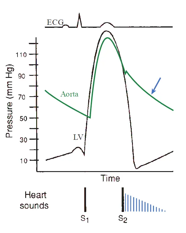
The following graph shows the hemodynamic profile of AR. During diastole, the aortic pressure falls rapidly, depicted by the arrow, and LV pressure rises as blood regurgitates from the aorta into the LV. A diastolic decrescendo murmur, beginning at S2, corresponds with the abnormal regurgitant flow.
Treatment
Clinical progression of patients with asymptomatic chronic AR and normal LV contractile function is very slow. Therefore, asymptomatic patients are monitored with periodic examinations, usually by serial echocardiography. Patients with asymptomatic severe AR may benefit from afterload reducing vasodilators (e.g. a calcium channel blocker or an ACE inhibitor) for treatment of accompanying hypertension. However, such agents do not prolong the compensated stage of chronic AR.
Symptomatic patients, or asymptomatic patients with severe AR and impaired LV contractile function (i.e. an ejection fraction ), should be offered surgical correction to prevent deterioration. Studies of such patients show that without surgery, death usually occurs within 4 years after the development of angina or 2 years after the onset of heart failure symptoms.
Tricuspid valve disease
Tricuspid Stenosis
Tricuspid stenosis (TS) is rare and is usually a long-term consequence of rheumatic fever. The OS and diastolic murmur of TS are similar to those of MS, but the murmur is heard closer to the sternum and it intensifies on inspiration because of increased right heart venous return.
In TS, the neck veins are distended and may show a large a wave as a result of right atrial contraction against the stenotic tricuspid valve orifice when sinus rhythm is present. Patients may develop abdominal distention and hepatomegaly owing to passive venous congestion. Percutaneous balloon dilatation or surgical correction (valvuloplasty or valve replacement) is usually required.
Tricuspid Regurgitation
Tricuspid regurgitation (TR) is usually functional rather than structural in nature; that is, it mostly results from right ventricular enlargement (e.g., owing to right-sided volume overload) rather than from primary valve disease.
Among patients with rheumatic mitral stenosis, 20% also have significant TR (of whom 80% have functional TR because of pulmonary hypertension with right ventricular enlargement, and 20% have structural TR resulting from rheumatic involvement of the tricuspid valve).
A rare cause of TR is carcinoid syndrome, in which a type of neuroendocrine tumor (usually from an origin in the gastrointestinal tract) releases serotonin metabolites into the bloodstream. These metabolites are thought to stimulate the formation of endocardial plaques in the right side of the heart. Involvement of the tricuspid valve immobilizes the leaflets, often resulting in substantial TR and, less often, TS.
The most common physical signs of TR are prominent v waves in the jugular veins and a pulsatile liver because of regurgitation of right ventricular blood into the systemic veins. The systolic murmur of TR is heard at the lower left sternal border.
Doppler echocardiography readily detects TR and can quantify it. The treatment of functional TR is directed at the conditions responsible for the elevated right ventricular size or pressure, and diuretic therapy; surgical repair of the valve is indicated in severe cases.
Pulmonic valve disease
Pulmonic Stenosis
Pulmonic stenosis (PS) is rare, and its cause is almost always congenital deformity of the valve. Carcinoid syndrome is another rare etiology, in which encasement and immobilization of the valve leaflets can occur.
The systolic crescendo–decrescendo murmur of PS is usually loudest at the second or third left intercostal space close to the sternum. It may radiate to the neck or left shoulder and is often preceded by an ejection click.
PS is considered to be severe if the peak systolic pressure gradient across the valve is mmHg, moderate if the gradient is 40 to 80 mmHg, and mild if the gradient mmHg. Only patients with moderate-to-severe gradients are symptomatic. In such cases, transcatheter balloon valvuloplasty is usually effective therapy.
Pulmonic Regurgitation
Pulmonic regurgitation (PR) most commonly develops in the setting of severe pulmonary hypertension and results from dilatation of the valve annulus by the enlarged pulmonary artery.
Auscultation reveals a high-pitched diastolic decrescendo murmur along the left sternal border that is often indistinguishable from AR. However, it can easily be distinguished from AR using Doppler echocardiography.
Prosthetic valves
Because all available valve substitutes have certain limitations, valve replacement surgery is not a true “cure.” Currently available valve substitutes include mechanical and bioprosthetic valves (derived from animal or human tissue) devices. One example of a mechanical valve is the St. Jude prosthesis, a hinged bileaflet valve consisting of two pyrolytic carbon discs that open opposite one another.
Mechanical valves, while durable, present foreign thrombogenic surfaces to the circulating blood and require life-long anticoagulation to prevent thromboembolism. In contrast, bioprosthetic valves display a very low rate of thromboembolism and do not require long-term anticoagulation therapy.
The most commonly used bioprostheses are made from glutaraldehyde-fixed porcine (pig) valves secured in a support frame. In addition, bovine (cow) pericardium and human homograft (aortic valves harvested and cryopreserved from cadavers) prostheses are used. For patients who undergo AVR because of endocarditis, human homograft replacements are especially useful because they have low rates of subsequent reinfection.
Bioprosthetic valves have limited durability compared with mechanical valves, and structural failure occurs in up to 50% by 15 years after implantation. The principal causes of failure are leaflet tears and calcification. Failure rates vary greatly depending on the position of the valve. For example, bioprosthetic valves in the mitral position deteriorate more rapidly than those in the aortic position. This is likely because the mitral valve is exposed to higher closing forces, resulting in greater leaflet stress.
Common to all types of prosthetic valves is the risk of infective endocarditis, which occurs with an incidence of 1% to 2% per patient per year. If endocarditis occurs in the first 60 days after surgery, the mortality rate is exceedingly high (50% to 80%). If endocarditis occurs later, mortality rates range from 20% to 50%. Reoperation is usually required when endocarditis involves a mechanical prosthesis because an adjacent abscess is frequently present. Some cases of bioprosthetic valve endocarditis may respond to antibiotic therapy alone.
The mortality and complication rates of mechanical and bioprosthetic valves are similar for the first 10 years after replacement. In 20-year follow-up studies of randomized, controlled trials, mechanical valves have been shown to be superior to bioprosthetic valves for event-free survival, except for bleeding complications related to anticoagulation therapy. Therefore, the decision about which type of prosthesis to use often depends on the patient’s expected lifespan in comparison to the longevity of the valve; risk-versus-benefit considerations of chronic anticoagulation therapy; and patient and surgeon preferences. Mechanical valves are often recommended for younger patients and for those who will be tolerant to and compliant with anticoagulant therapy. Bioprosthetic valves are generally suitable choices for patients 65 years of age or older and for patients with contraindications to chronic anticoagulation.
See also
References
Additional Reading
- Fowler, V. G., Durack, D. T., Selton-Suty, C., et al. (2023). The 2023 Duke-International Society for Cardiovascular Infectious Diseases Criteria for Infective Endocarditis: Updating the Modified Duke Criteria. Clinical Infectious Diseases, 77(4), 518–526. https://doi.org/10.1093/cid/ciad271
- Gerber MA, Baltimore RS, Eaton CB, et al. Prevention of rheumatic fever and diagnosis and treatment of acute streptococcal pharyngitis. Circulation. 2009;119:1541–1551
- Habib G, Hoen B, Tomos P, et al. Guidelines on the prevention, diagnosis, and treatment of infective endocarditis (new version 2009); The Task Force on the Prevention, Diagnosis, and Treatment of Infective Endocarditis of the European Society of Cardiology. Eur Heart J. 2009;30:2369–2413
- Nishimura RA, Otto CM, Bonow RO, et al. 2014 AHA/ ACC guideline for the management of patients with valvular heart disease: a report of the American College of Cardiology/ American Heart Association Task Force on Practice Guidelines. J Am Coll Cardiol. 2014;63:e57-e185
- O’Gara P, Loscalzo J. Valvular heart disease. In: Longo DL, Fauci AS, Kasper DL, et al., eds. Harrison’s Principles of Internal Medicine. 18th ed. New York, NY: McGraw-Hill; 2012:1929–1950
- Vahanian A, Alferi O, Andreotti F, et al. Guidelines on the management of valvular heart disease (version 2012); The Joint Task Force on the Management of Valvular Heart Disease of the European Society of Cardiology and the European Association for Cardio-Thoracic Surgery. Eur Heart J. 2012;33:2451–2496
- Wilson W, Taubert KA, Gewitz M, et al. Prevention of infective endocarditis: guidelines from the American Heart Association: a guideline from the American Heart Association Rheumatic Fever, Endocarditis, and Kawasaki Disease Committee, Council on Cardiovascular Disease in the Young, and the Council on Clinical Cardiology, Council on Cardiovascular Surgery and Anesthesia, and the Quality of Care and Outcomes Research Interdisciplinary Working Group. Circulation. 2007;116:1736–1754